Caffeine's effects on resting-state oxygen and glucose metabolism: A combined MR and PET study Y. Chen1, A. B. Newberg2, J. wang1, H. Rao1, H. An3, J. Greenberg4, N. Wintering2, V. Tolles2, and J. A. Detre1
1Center of Functional Neuroimaging, University of Pennsylvania, Philadelphia, PA, United States, 2Department of Radiology, University of Pennsylvania, Philadelphia,
PA, United States, 3Department of Radiology, University of North Carolina, Chapel Hill, NC, United States, 4Department of Neurology, University of Pennsylvania,
Introduction Caffeine is part of the methylxanthine family, which are adenosine antagonists [1]. Since adenosine mediates both blood flow and neural activity, it has been suggested that caffeine has the ability to alter the tight coupling between blood flow and oxygen metabolism. However, most studies to-date have focused on caffeine’s effects on functional activations relative to baseline [2,3]. In this study, we examine caffeine’s effects on resting-state cerebral glucose and oxygen metabolism using a combination of MRI and PET. Methods MR Imaging: Data from five healthy subjects were collected on a Siemens 3T MR system (Siemens Trio, Erlangen, Germany). Subjects were requested to abstain from caffeine for 12 hours prior to the study. Each experiment consisted of two identical sessions, between which subjects waited outside the scanner for 2-3 hours. In the middle of the intersession period, subjects ingested an over-the-counter caffeine pill (200mg of caffeine). In each of the sessions, cerebral blood flow (CBF) images were acquired using a continuous arterial spin labeling (ASL) tagging scheme with gradient EPI readout (TR=4s, TE=17ms, post labeling delay=1.2s, 70 pairs of interleaved control and tag images). A double-echo asymmetric echo spin echo EPI sequence [4] was used to calculate maps of R2’ and oxygen extraction fraction
(OEF). Cerebral metabolic rate of oxygen (CMRO2) maps were calculated as the product of CBF and OEF maps. During all scans, subjects were instructed to close
their eyes in order to maintain resting-state. A high resolution T1-weighted anatomical image was also collected. PET Imaging: At the beginning of each ASL scan, approximately 0.14 mCi/kg of 18F-labeled fluorodeoxyglucose (FDG) was injected through an intravenous catheter to allow for FDG uptake during the ASL scan. Arterial plasma samples were obtained at increasing time intervals following the FDG administration and analyzed for 18F activity and glucose level, permitting quantification of cerebral metabolic rate of glucose (CMRGlu) [5]. PET images were acquired on a G-PET brain scanner [6] immediately following each MR session and at least 45 minutes after FDG administration. Data Analysis: CBF, CMRO2, OEF, CMRGlu and CMRO2: CMRGlu from all subjects were obtained from 8 regions of interest (ROI), which include: anterior cingulate,
amygdala, calcarine cortex, caudate, insula, orbital frontal cortex, superior parietal cortex and thalamus. Pre- and post-caffeine data were compared using repeated measures Analysis-of-variance (ANOVA) (SPSS v.17, SPSS Inc., Chicago, IL).
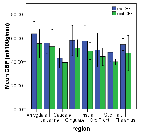
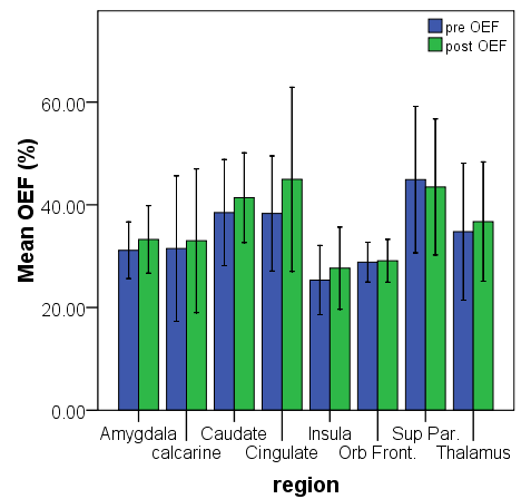
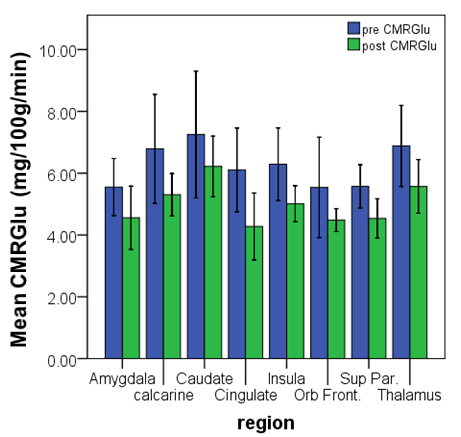
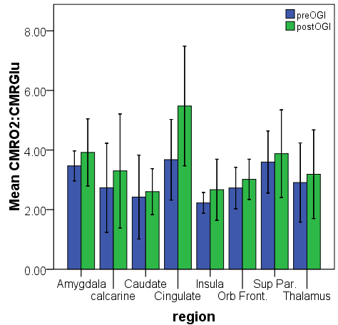
Fig. 1 Plots of mean (± 95% CI) pre- and post-caffeine CBF, OEF, CMRGlu and CMRO2: CMRGlu (OGI).
Results Plots of mean pre- and post-caffeine CBF, OEF, CMRGlu and CMRO2: CMRGlu are shown in Fig. 1. ANOVA results
show that caffeine decreases CBF (mean change=-10%, F=13.819, p=0.001) and CMRGlu (mean change=-18%, F=36.41, p<<0.05) and increases OEF (mean change=+7%, F=8.571, p=0.006) and the CMRO2: CMRGlu ratio (mean
change=+22%, F=10.725, p=0.003) in all regions. Caffeine has no effect on CMRO2. Post-hoc Tukey’s HSD shows that
caudate and anterior cingulate have significantly different CBF (p=0.023), CMRGlu (p=0.017) and CMRO2: CMRGlu
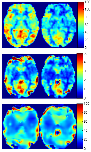
(p=0.014). Fig. 2 shows sample CBF, CMRO2 and OEF maps obtained from the MR scans of a representative subject. Discussion Caffeine’s vasoconstrictive effects have been well documented [7]. Given that CMRO2∝OEF⋅CBF, the decrease in CBF
in the presence of caffeine suggests OEF should increase in order to maintain CMRO2. This was indeed observed in the
current study. These results also provide a better understanding of the decrease in baseline BOLD signal associated with caffeine: the decreased CBF accompanied by the increased OEF results in an increase in local deoxyhemoglobin concentration, which in turn causes a decrease in the MR signal. An interesting finding in this study is that caffeine appears to alter the balance of oxygen and glucose metabolism during resting-state, and this change is particularly prominent in the anterior cingulate. Since this region has been shown to be associated with wakefulness and sleep [8], the altered CMRO2: CMRGlu ratio could be the mechanism through which
caffeine affects alertness. References 1. Fredholm BB et al. Pharmacol Rev. 51(1) 83, 1999. 2. Mulderink TA et al. Neuroimage 15(1):37, 2002. 3. Koppelstaetter, Neuroimage 31(S1), 438, 2006. 4. An & Lin Magn Reson Med. 50(4):708 2003. 5. Reivich M et al. Circ Res. 44: 127 1979. 6. Karp JS et al. J Nucl Med. 44(8):1240 2003. 7. Field et al. Radiology 227:129 2003. 8. Maquet P. J.
Proc. Intl. Soc. Mag. Reson. Med. 17 (2009)
RECENT DEVELOPMENTS Product Liability—Eighth Circuit Adopts Narrow View: Only APLA Failure-to-Warn/Mislabeling Claims Against Generic Manufacturers Are Preempted Under Mensing ; Design- Defect and Implied-Warranty Claims Are Still Viable The Court of Appeals for the Eighth Circuit recently issued a pair of opinions interpreting the United States Supreme Court’s decisions in
Klinik für Chirurgie Kantonsspital St.Gallen Sakrale Nervenstimulation – Teststimulation, Implantation Testelektrode Teststimulation (= periphere Nervenevaluation) durchgeführt. Dieser Eingriff erfolgt ambulant. Begriffserklärung Stuhlinkontinenz wird unter anderem durch Diese Operation wird in örtlicher Betäubung durch ein gestörtes Empfinden des Enddarmes Op
 Caffeine's effects on resting-state oxygen and glucose metabolism: A combined MR and PET study