Photos.or.kr

Journal of Photoscience (2001), Vol. 8(1), pp. 1−7 Photosynthetic Response and Protective Regulation To Ultraviolet-B
Radiation In Green Pepper (Capsicum annuum L.) Leaves
Dae Whan Kim1, Sung-Soo Jun and Young-Nam Hong School of Biological Sciences, Seoul National University, Seoul 151-742, Korea Kumho Life and Environmental Science Laboratory, Kwangju 500-712, Korea The deteriorative effect of ultraviolet-B (UV-B) radiation on photosynthesis was assessed by the simultaneousmeasurement of O evolution and chlorophyll (Chl) fluorescence in green pepper. UV-B was given at the intensity of 1 W·m-2, a dosage often encountered in urban area of Seoul in Korea, to the detached leaves. Both Pmax andquantum yield of O evolution was rapidly decreased, in a parallel phase, with increasing time of UV-B treatment.
Chl fluorescence parameters were also significantly affected. Fo was increased while both Fm and Fv weredecreased. Photochemical efficiency of PS II (Fv / Fm) was also declined, although to a lesser extent than Pmax.
Both qP and NPQ were decreased similarly with increasing time of UV-B treatment. However, PS I remainedstable. The addition of lincomycin prior to UV-B treatment accelerated the decline in Fv / Fm to some extent,suggesting that D1 protein turnover may play a role in overcoming the harmful effect of UV-B. The amount ofphotosynthetic pigments was less affected than photosynthetic response in showing decline in Chl a and carotenoidsafter 24 h-treatment. Presumptive flavonoid contents, measured by changes in absorbance at 270 nm, 300 nm and330 nm, were all increased by roughly 50% after 8 h-treatment. Among antioxidant enzymes, activities of catalaseand peroxidase were steadily increased until 12 h of UV-B treatment whereas ascorbate peroxidase, dehydroascorbatereductase and glutathione reductase did not show any significant change. The results indicate that deteriorativeeffect of UV-B on photosynthesis precedes the protection exerted by pigment synthesis and antioxidant enzymes.
key words: UV-B radiation, pepper, photosynthesis, chlorophyll fluorescence, flavonoid, antioxidant enzyme
INTRODUCTION
absorbing biomolecules, namely proteins and nucleic acidswere photomodified [9,10]. In addition, UV-B radiation resulted Plants in nature are continuously affected by various natural in changes in pigment composition by reducing Chl content environmental factors such as light, water and temperature and increasing flavonoid content, which was especially and respond to changes in those factors to their advantage.
After rapid industrialization, anthropogenic environmental Decrease in photosynthesis due to UV-B radiation was pollutants such as heavy metals and airborne pollutants presented mainly attributed to the inactivation of PS II [12-15]. UV-B a new set of adverse factors directly or indirectly affecting treated leaves showed reduction in O evolution and Chl plants growth. Among these factors, UV-B (between 280 and fluorescence originating from PS II [15]. The same results 320 nm) has drawn attention in recent years since the heavy were observed in the isolated chloroplasts from UV-B treated usage of Freon (chloroflurocarbon) gas for aerosol propellants plants [12]. UV-B mostly affected PS II reaction center itself and refrigeration resulted in the rapid erosion of stratospheric and water oxidizing complex [16,17]. The outcome was due ozone layer on a global scale [1,2]. In consequence, UV-B to the denaturation of proteins by UV-B in PS II complex, level reaching on the ground has greatly increased [3]. For especially in D1 protein and LHCP II and subsequent damage example, UV-B radiation was in the range of 0.9 W· m-2 during in Q was followed [13,17]. D2 protein, generally thought to the midday in Seoul according to a recent report of Korea be unaffected, was also shown harmed [18]. Destruction of Trp residue in the ATPase protein resulted in the inactivation Increased UV-B radiation was shown to affect harmfully of ATPase [19]. UV-B reduced RNA transcription of cab and most physiological and biochemical processes in plants including psb A [20]. On the other hand, PS I and cytochrome b-f photosynthesis [4,5], dark respiration [4,5], transpiration [5], complex were hardly affected by UV-B [15].
biomass allocation [6], and leaf expansion [7,8]. UV-B UV-B affected the dark reaction as well by reducing irradiation could also be damaging when essential UV- Rubisco activities [15,20]. Both large (55 kDa) and small (15kDa) subunits of Rubisco were degraded and synthesis of *To whom correspondence should be addressed.
their mRNA was declined after long exposure to UV-B [20].
The photomodification of Trp residue by UV-B in the large Received 10 December 2000; accepted 25 January 2001 subunit produced 66 kDa subunit instead of 55 kDa subunit [21]. All of them led to the decline in Rubisco activities and subsequent reduction in CO assimilation.
Measurement of O evolution and Chl fluorescence. Chl UV-B was absorbed directly by DNA to form UV-induced fluorescence and O evolution were measured simultaneously photoproducts such as cyclobutane pyrimidine dimer and using leaf discs of 3.5 cm-diameter, taken from leaves of randomly pyrimidine (6,4) pyrimidone dimer, which caused serious selected plants, in Hansatech (Kings Lynn, UK) LD2 leaf disc errors in replication and transcription [9,22]. To ameliorate chamber. After UV-B treatment, leaves were dark-adapted for 30 these deteriorative effects, plants developed two types of min in a floating state on DW. Each leaf disc was chosen to have protective mechanisms: DNA repair and shielding to reduce approximately equal Chl content, when measured by Minolta Chl DNA damage. Photolyase was shown to be involved in repairing meter (SPAD-502, Minolta, Japan) and normalized to have equal UV-B damaged DNA in plants [23, 24]. Thickening of leaves Chl content. The measurement of Chl content by SPAD-502 was and formation of cuticular waxes were manifested to prevent reliably matched with that of conventional extraction method the penetration of UV-B [25]. Accumulation of proline, ferulic using acetone. Chl fluorescence was measured using Walz PAM acid, and flavonoids was observed in the leaves [26,27]. Among Chl fluorometer (Effeltrich, Germany) while O evolution was them UV-absorbing flavonoids and transcripts involved in polarographically measured with a Clark type electrode at 25oC flavonoid biosynthesis were increased after long exposure to using Hansatech O electrode control box. To measure Pmax and UV-B [27-29]. Increase in flavonoid content decreased the quantum yield of O evolution, actinic light was given at three UV-induced dimer formation [30]. Furthermore, UV-B radiation different intensities of 30, 50, and 270 µmol · m-2· s-1 using Schott led to the formation of biologically active oxygenic species illuminator (Schott Glass, Stafford, UK). Fv/Fm, NPQ (defined that could damage the photosynthetic apparatus and adversely as Fm/Fm’-1), and qP were calculated as described before [34].
affect the enzyme activities. Antioxidant enzymes such as PS I oxidation and reduction kinetics was measured by monitoring catalase, ascorbate peroxidase, and glutathione peroxidase changes in absorbance at 820 nm (∆A ) using PAM Chl were activated against oxidative stresses [31]. Accordingly, fluorometer connected to 830 nm LED (type ED 800T) as activities of various antioxidant genes and enzymes were increased after UV-B treatment [32,33].
Lincomycin treatment. Lincomycin was treated by immersing In the present study, the multiple effects of UV-B and plants’ the petiole of each detached leaf in a Eppendorf tube containing response against it were studied in green pepper, a major 1.2 mM lincomycin solution for 4 h in the growth chamber. UV-B produce in Korea. We first attempted to evaluate the deteriorative treatment was subsequently done to the lincomycin-imbibed leaves.
effect of realistic UV-B dosage encountered often in Korea, Determination of photosynthetic pigments. The photosynthetic on photosynthesis. Next the lesion sites were identified and pigments were extracted following the method of Hiscox and assessed by indices of Chl fluorescence parameters. Finally, Israelstham [36]. In 15 mL tube, leaf segments were put with 5 the physiological protective responses in terms of pigment mL of DMSO and incubated for 1 h at 65°C. The extracted composition and regulation of antioxidant enzymes were solution was diluted twice with the same volume of DMSO and absorbance was read at 470, 645, and 663 nm, respectively. Theamount of Chl a, Chl b, and total carotenoids was calculatedaccording to the formula of Lichtenthaler [37] as given below.
MATERIALS AND METHODS
Plant material. Green pepper plants (Capsicum annuum L. cv.
Saemaeul Kumjang #3) were grown in pots filled with soil(Bioplug #2, Hungnong Seeds Co, Ltd., Korea) for 4 to 5 weeks Total carotenoid = (1000 A − 1.82 Chl a − 85.02 Chl b) / 198 in a growth chamber maintained at 25 ± 1oC under the photoperiodof 16 h-light and 8 h-dark. Light was provided by True-lite II Measurement of UV-B absorbing pigments. The amount of fluorescent lamps (Durotest, USA) at the intensity of 200 UV-B absorbing flavonoids was estimated according to Krizek µmol ·m-2·s-1. Randomly selected leaves of similar size were et al. [38]. Leaf slices were extracted in 15 mL of solution sampled for subsequent UV-B treatment.
containing 40% (V/V) methanol and 1% (V/V) HCl and boiled UV-B treatment. UV-B, at the intensity of 1 W · m-2 was for 3 min in a water bath. The extracted solution was left at RT directly cast on the detached leaves floating on DW at RT. The for 24 h and the absorbance values at 270 nm, 300 nm, and 330 intensity of UV-B was adjusted to be slightly higher than the nm were monitored. The absorbance values were calculated daily maximal dosage (0.9 W ·m-2) in urban area of Seoul in May.
UV-B radiation was provided by the additional attachment of a Activities of antioxidant enzymes. To measure the activities of UV-B lamp (XX-15B, Spectronics Corporation, USA), covered antioxidant enzymes, crude extract was obtained by grinding 1 g with cellulose acetate membrane to eliminate UV below 280 nm, of leaf slices with 2 mL of solution containing 85 mM K-PO4 to the fluorescent lamps in the growth chamber. UV-B intensity buffer (pH 7.8) and 1% (W/V) polyvinylpyrrolidone in a mortar was determined by Digital Radiometer (DRC-100X, Spectronics and centrifuging at 18000 g for 25 min. CAT, GR, and POD were Photosynthetic Response and Protective Regulation to UV-B Radiation assayed according to Rao et al. [32]. CAT activity was determinedspectrophotometrically by the decrease of absorbance at 240 nmin a cuvette with 1 mL of reaction mixture containing 100 mMK-PO (pH 7.0), 40 µL of crude extract, and 10 mM H O , which was added just prior to the initiation of reaction. GR was assayedin 1 mL of reaction mixture containing 100 mM K-PO (pH 7.8), 0.2 mM NADPH, 0.5 mM GSSG (oxidized glutathione), 2 mMEDTA, and 100 µL of crude extract by monitoring decrease ofabsorbance at 340 nm. The assay was initiated by the addition ofNADPH. POD activity was quantified by measuring the rate ofguajacol tetramerization shown as the increase in absorbance at470 nm in a cuvette containing 1 mL of reaction mixture of 100mM K-PO (pH 6.5), 16 mM guajacol, 10 mM H O , and 100 µL of crude extract. The reaction was initiated by adding crudeextract and followed for 10 min.
APX and DHAR were measured by following the method of Asada [39]. APX was monitored by the decrease in absorbance at Figure 1. Changes in the Pmax and quantum yield of O evolution in the UV-B treated leaves. Data presented are mean values ± S.E.
290 nm in 1 mL of reaction mixture composed of 50 mM K-PO4 (pH 7.0), 0.5 mM ascorbate, 0.1 mM H O , and 5 µL of crude extract. DHAR was assayed in 1 mL of reaction mixture containing50 mM K-PO (pH 6.5), 0.5 mM dehydroascorbate, 5 mM GSH yield implies that UV-B treatment may not make green pepper (reduced glutathione), 0.1 mM EDTA, and 15 µL of crude extract more susceptible to photoinhibition. Pmax was decreased more by monitoring increase of absorbance at 256 nm.
rapidly than quantum yield after UV-B treatment in coleus [40]. Changes in Chl fluorescence parameters RESULTS AND DISCUSSION
The initial fluorescence (Fo) and maximal photochemical efficiency (Fv/Fm) are general indicators for PS II functionality.
General effect of UV-B on leaf morphology Damages to PS II are often accompanied with rise in Fo level The detached leaves did not show any significant change in and drop in Fv/Fm. Changes in Fo reflect structural alterations appearance after 24 h of UV-B treatment. However, the leaves at the antenna of PS II while those in Fv/Fm indicate varied became more darkened and glossy after a few h of UV-B energy capturing efficiency in PS II [41]. As shown in Fig.
treatment. Morphological alteration such as thickening of 2A, after UV-B treatment Fo was increased whereas both Fm leaves and formation of cuticular waxes were shown to be the and Fv were decreased rapidly in the initial 2 h. Photochemical primary response to UV-B in terrestrial plants [25]. In green efficiency of PS II, represented by Fv/Fm, was linearly declined, pepper, the leaves also became glossy and dark, possibly due although to a lesser extent than Pmax, showing 12%, 23%, 29% to the accumulation of waxes and some UV-B absorbing and 39% reduction after 1, 2, 3 and 4 h of UV-B treatment (Fig. 2B). The linear decrease in Fv/Fm was also observed inDunaliella after UV-B treatment [42].
Effect of UV-B on Pmax and quantum yield of O evolution After onset of illumination, the maximal Chl fluorescence To evaluate the extent of deteriorative effect of UV-B on the declines as photosynthetic reaction proceeds due to the photosynthetic capacity, Pmax and quantum yield of O photosynthetic electron flow represented as qP, and nonradiative evolution were measured after direct treatment of UV-B on energy dissipation and ∆pH formation manifested as NPQ the detached leaves. Pmax was determined under saturating [34]. Both qP and NPQ were decreased linearly in a parallel PFR (270 µmol· m-2·s-1) while quantum yield was determined phase by the increased time of UV-B treatment (Fig. 3).
under low PFR (30 and 50 µmol· m-2· s-1). As shown in Fig. 1, Decreased qP indicates that the acceptor side of PS II (Q ) is Pmax was decreased about 28% after 1 h, 47% after 2h, 49% overly reduced by hindered electron flow [34]. NPQ usually after 3 h, and 64% after 4 h while quantum yield was decreased increases when qP is decreased [44]. Decreased NPQ about 18%, 42%, 46%, and 59%, respectively. Although accompanied with decreased qP may reflect the damage in decrease in Pmax preceded decrease in quantum yield, both ATPase by UV-B, which would block DpH formation by Pmax and quantum yield were linearly decreased in a parallel increased back pressure. APTase was shown harmed in earlier phase in relation to the increased time of UV-B treatment, demonstrating both maximal photosynthetic activity and Changes in absorbance at 820 nm reflects the oxidation and efficiency in the photosynthetic apparatus were similarly reduction state of P-700 that reflects electron flow from PS II affected by UV-B. Parallel decrease in Pmax and quantum to PS I and from PS I to NADP+ [43]. After 4 h of UV-B 
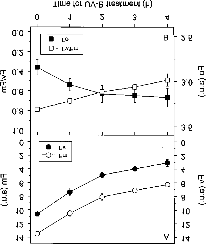
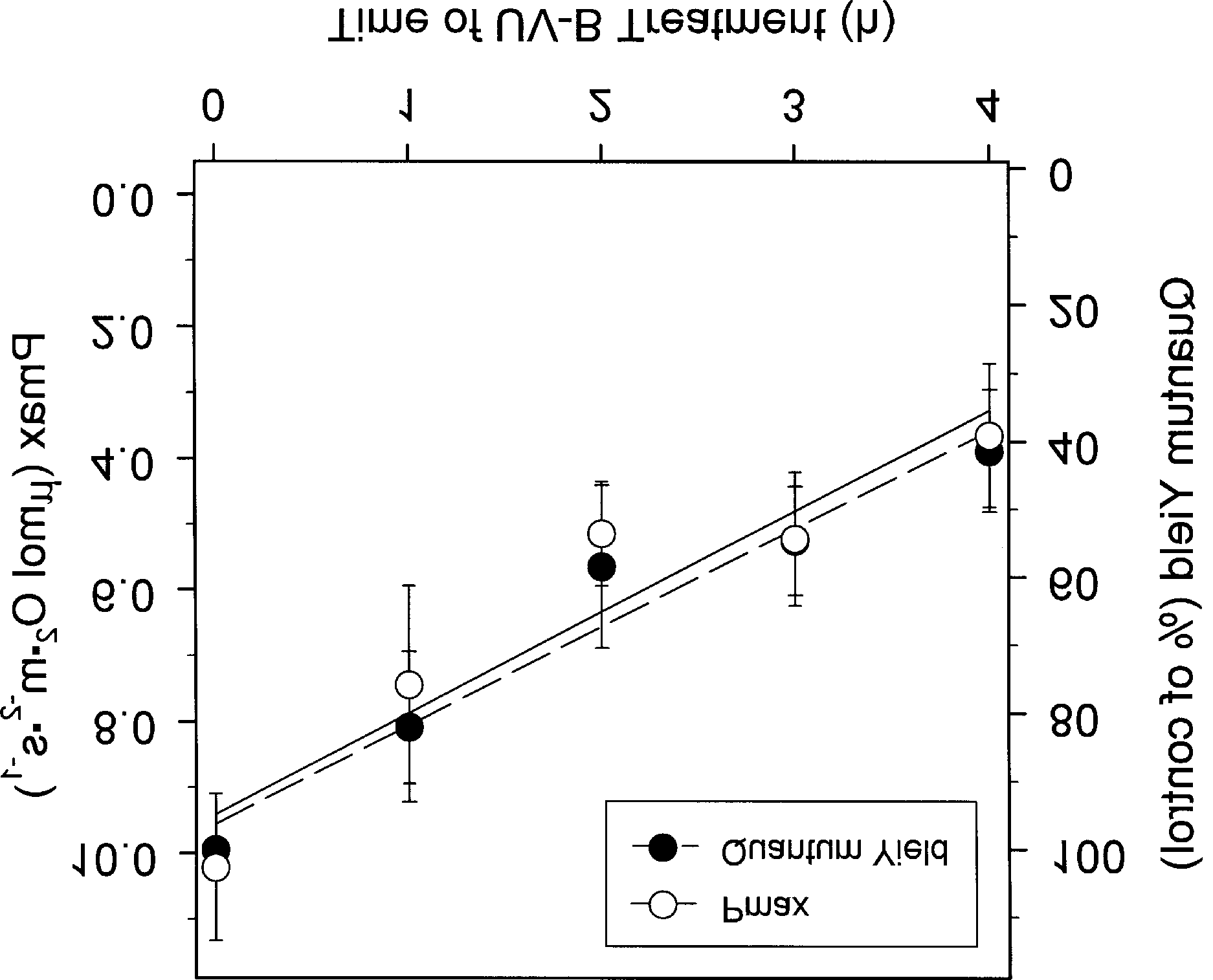
Figure 2. Changes in various Chl fluorescence parameters in theUV-B treated leaves: (A) the maximal (Fm) and variable (Fv)fluorescence, (B) the initial (Fo) and the maximal photochemicalefficiency of PSII (Fv/Fm). Fo, Fm, and Fv are given in arbitraryunits. Data presented are mean values ± S.E. for 5 measurements.
Figure 4. Changes in the Pmax (A), Fv/Fm (B), and Fo (C) in thecontrol and lincomycin-treated leaves after UV-B treatment. Datapresented are mean values ± S.E. for 5 measurements.
effect in photoinhibition. Similarly, UV-B treatment damagedD1 proteins [17]. In view of these, UV-B was treated to thelincomycin-infiltrated leaves to test the role of D1 proteinturnover in alleviating the adverse effect of UV-B. As shownin Fig. 4A, decrease in Pmax was identical in control andlincomycin-treated leaves. However, decrease in Fv/Fm andincrease in Fo was accelerated in lincomycin-treated leaves(Fig. 4B and 4C). The decrease in qP and NPQ was alsoaccelerated in lincomycin-treated leaves (Fig. 3). The results Figure 3. Changes in the photochemical quenching (qP) and implicate that D1 protein turnover would play a role in nonphotochemical quenching (NPQ) in the control and lincomycin- treated leaves after UV-B treatment. Data presented are mean values Using antisensor lines of Arabidopsis plants with reduced content in xanthophyll, the effect of UV-B treatment was alsotested. Antisensor plants showed more rapid decline in Fv/ treatment absorbance at 820 nm was not changed in P-700 Fm. In addition, decrease in qQ and NPQ occurred faster oxidation-reduction kinetics (data not shown). Therefore, PS I (data not shown). These results indicate that xanthophyll appeared to be resistant to UV-B damage as previously cycle is also involved in relieving the deteriorative effect of UV-B. Therefore, photoprotective mechanisms appear to bebeneficiary for reducing the adverse effect of UV-B.
Effect of lincomycinTurnover of D1 protein plays a significant role in overcoming Effect on the contents of photosynthetic pigments photoinhibition as lincomycin-infiltrated leaves show exacerbating The adverse effect of UV-B on photosynthesis may result in Photosynthetic Response and Protective Regulation to UV-B Radiation Table 1. Changes in the amount of photosynthetic pigments afterUV-B treatment.
*Data presented are mean values ± S.E. for 5 measurements.
part from the declined photosynthetic pigments as UV-B wasshown to degrade Chl. As shown in Table 1, UV-B irradiancehad negligible effect on the content of photosynthetic pigments Figure 5. Changes in the contents of UV-B absorbing flavonoids up to 12 h of treatment. Total amounts in Chl a, Chl b, and after UV-B treatment. Changes in flavonoid contents were estimated carotenoids all remained unchanged. At this time point the by measuring the absorbance values at 270, 300, and 330 nm of photosynthesis presumably stops completely. Thus it appears pigment solution extracted in ethanol. Data presented are mean that initial decline in photosynthesis after UV-B treatment is independent of contents in photosynthetic pigments. However,after 24 h of treatment Chl a was declined about 25% and Effect on the activities of antioxidant enzymes carotenoids were decreased about 40% (Table 1). In contrast, Under UV-B radiation, high amounts of free radicals are Chl b stayed little changed. In Pisum sativum, decrease in Chl produced, generating, in turn, active oxygenic species such as a and b accompanied with formation of chlorophyllides a and H O although the exact mechanism by which they are b was observed [44]. Decrease in carotenoids was contradictory generated is unknown. Under such oxidative stresses, plants to the result in Pisum sativum in which carotenoid content invoke the antioxidant defense systems [29,31]. One consists was increased relative to the Chl content [44]. The increased of low molecular weight antioxidants such as ascorbate, carotenoids provide photoprotection under high light from glutathione, and carotenoids. The other is composed of various photoinhibitory damage. In our experiment, UV-B was sup antioxidant enzymes that are activated to scavenge the potentially plemented with relatively low light (200 µmol· m-2· s-1) where dangerous active species [32,33]. We tested 5 representative photoinhibition was presumably lacking. Our results implicate antioxidant enzymes, namely APX, CAT, DHAR, GR, and that UV-B per se may photooxidize carotenoids to some POD under UV-B treatment. Activities of CAT and POD extent, resulting in their degradation.
Effect on the contents of UV-B absorbing pigmentsPlants protect themselves against UV light by accumulating screening pigments. Colorless flavonoids were accumulatedin the epidermal tissues of UV-treated plants and reduceddamage from UV radiation [27,28]. In view of this, changesin presumptive flavonoid contents after UV-B treatment wereinvestigated by monitoring changes in absorbance values at270, 300, and 330 nm. As shown in Fig. 5, absorbance valuesat three wavelengths did not change until 4 h after UV-Btreatment. However, on the 8th h, the absorbance values at allwavelengths were roughly 50% increased and stayed on the samelevel on the 12th h. The induction of flavonoid accumulationseemed to occur far after significant decline of photosynthesis,which implied that flavonoid accumulation would not helpprotecting the photosynthetic apparatus. Time course ofinduction was correlated with transcription of enzymes involved Figure 6. Changes in the activities of various antioxidant enzymes in flavonoid synthesis [29]. Transcription of chalcone synthase after UV-B treatment: CAT, catalase; DHAR, dehydroascorbate and 4-coumaroyl-CoA ligase reached to maximal level after 6 reductase; POD, peroxidase. Data presented are mean values ± S.E.
increased with increasing time of UV-B treatment whereas D. Menzies, M. Ondrusek, Z. Wan and K. J. Waters (1992) that of DHAR did not change (Fig. 6). POD activity was Ozone depletion: ultraviolet radiation and phytoplankton induced more rapidly and in a higher degree. APX showed a biology in Antarctic water. Science 255, 952-959.
similar tendency as DHAR, but GR did not exhibit any 4. Sisson, W. B. and M. M. Caldwell (1976) Photosynthesis, consistent trend (data not shown). The H O scavenging system dark respiration, and growth of Rumex patientia L. exposed composed of GR, DHAR, and APX (ascorbate-glutathione to ultraviolet irradiance (288-315 nm) simulating a reduced
ozone column. Plant Physiol. 58, 563-568.
cycle) did not seem to be a main system to remove active 5. Teramura, A. H., R. H. Biggs and S. Kossuth (1980) Effect oxygenic species generated by UV-B in green pepper. It was of ultraviolet-B irradiance on soybean. II. Interaction previously shown that enzymes of the ascorbate-glutathione between ultraviolet-B and photosynthetically active radiation cycle were less affected by UV-B than by ozone [32].
on net photosynthesis, dark respiration, and transpiration.
Plant Physiol. 65, 483-488.
6. Teramura, A. H. (1980) Effect of ultraviolet-B irradiance on CONCLUSION
soybean. I. Importance of photosynthetically active radiationin evaluating ultraviolet-B irradiance effects on soybean and Treatment of UV-B radiation, on a dosage often encountered in wheat growth. Physiol. Plant. 48, 333-339.
Korea, to the detached leaves of green pepper led to the rapid 7. Sisson, W. B. and M. M. Caldwell (1977) Atmospheric reduction in photosynthesis manifested as parallel drop in both ozone depletion: reduction of photosynthesis and growth of a Pmax and quantum yield of O evolution. Chl fluorescence sensitive higher plant exposed to enhanced UV-B radiation.
parameters were similarly affected to bring in the increase in J. Exp. Bot. 28, 691-705.
Fo, but decrease in Fm, Fv, and Fv/Fm. Both qP and NPQ 8. Dickson, J. G. and M. M. Caldwell (1978) Leaf development were decreased after UV-B treatment. The results would of Rumex patientia L. (Polygonaceae) exposed to UV
irradiation (280-320 nm). Am. J. Bot. 65, 857-863.
indicate that UV-B primarily affected PS II, but other sites 9. Quaite, F. E., B. M. Sutherland and J. C. Sutherland (1992) were also affected as shown by decrease in both qP and NPQ.
Action spectrum for DNA damage in alfalfa lowers However, PS I remained unaffected. D1 protein turnover and predicted impact of ozone depletion. Nature 358, 576-578.
xanthophylls cycle played a role in alleviating the UV-B induced 10. Caldwell, C. R. (1993) Ultraviolet-induced photodegradaion inhibition as in photoinhibition. The Chl a and carotenoids of cucumber (Cucumis sativus L.) microsomal and soluble were declined far after the decline of the photosynthesis.
protein tryptophanyl residues in vitro. Plant Physiol. 101,
Flavonoid contents and activities of some antioxidant enzymes were increased, but in a slower pace than reduction in 11. Mirecki, R. M. and A. L. Teramura (1984) Effect of photosynthesis. Our results confirm that earlier reports showing ultravioet-B irradiance on soybean. V. The dependence of that UV-B was deteriorative on photosynthesis, especially in plant sensitivity on the photosynthetic photon flux density PS II, and invoked the flavonoid synthesis and activated during and after leaf expansion. Plant Physiol. 74, 475-480.
antioxidant enzymes, are valid in green pepper. The results 12. Iwanzik, W., M. Tevini, G. Dohnt, M. Voss, W. Weiss, P.
would also suggest that under UV-B the photosynthetic Gräber and G. Renger (1983) Action of UV-B radiation on apparatus is rapidly damaged but the protective mechanisms photosynthetic primary reactions in spinach chloroplasts.
including synthesis of flavonoids to shield UV and antioxidant Physiol. Plant. 58, 401-407.
enzymes to remove active oxygenic species are set to operate 13. Greenbreg, B. M., V. Gaba, O. Canaani, S. Malkin, A. K.
Mattoo and M. Edelman (1989) Separate photosystem II
reaction center protein in the visible and UV spectral
regions. Proc. Natl. Acad. Sci. USA 88, 6617-6620.
14. Renger, G., M. Volker, H. J. Eckert, R. Fromme, S. Hohm- Acknowledgement − This work was supported by S.N.U. Research Veit, and P. Gräber (1989) On the mechanism of photosystem II deterioration by UV-B irradiation. Photochem. Photobiol.
49, 97-105.
15. Strid, Å., W. S. Chew and J. M. Anderson (1990) Effects of REFERENCES
supplementary ultraviolet-B radiation on photosynthesis in
Pisum sativum. Biochim. Biophys. Acta 1020, 260-268.
1. Milina, J. J. and F. S. Rowland (1974) Stratospheric sink for 16. Vass, I., L. Sass, C. Spetea, A. Bakou, D. F. Ghanotakis and chlorofluoromethanes; chlorine atom-catalysed destruction V. Petrouleas (1996) UV-B-induced inhibition of photosystem of ozone. Nature 249, 810-812.
II electron transport studied by EPR and chlorophyll 2. Anderson, J. G., D. W. Toohey and W. H. Brune (1991) Free fluorescence. Impairment of donor and acceptor side radicals within the Antarctic vortex: The role of CFCs in components. Biochemistry 35, 8964-8973.
Antarctic ozone loss. Science 251, 39-46.
17. Melis, A., J. A. Nemson and M. A. Harrison (1992) Damage 3. Smith, R. C., B. Prezelin, K. S. Baker, R. R. Bidigare, N. P.
to functional components and partial degradation of Boucher, T. Coley, D. Karentz, S. Macintyre, H. A. Matlick, photosystem II reaction center proteins upon chloroplast Photosynthetic Response and Protective Regulation to UV-B Radiation exposure to ultraviolet-B radiation. Biochim. Biophys. Acta 17, 507-523.
1100, 312-320.
32. Rao, M. V., G. Paliyath and D. Ormrod (1996) Ultraviolet-B- 18. Jensen, M. A. K., V. Gaba, B. M. Greenberg, A. K. Mattoo and ozone-induced biochemical changes in antioxidant and M. Edelman (1996) Low threshold levels of ultraviolet- enzymes of Arabidopsis thaliana. Plant Physiol. 110, 125-
B in a background of photosynthetically active radiation trigger rapid degradation of the D2 protein of photosystem- 33. Willekens, H., W. V. Camp, M. V. Montagu, D. Inze, C.
II. Plant J. 9, 693-699.
Langebartels and H. Sandermann, Jr. (1994) Ozone, sulful 19. Imbrie, C. W. and T. M. Murphy (1984) Mechanism of dioxide, and ultraviolet B have similar effects on mRNA photoinactivation of plant plasma membrane ATPase.
accumulation of antioxidant genes in Nicotiana lumbaginifolia Photochem. Photobiol. 40, 243-248.
L. Plant Physiol. 106, 1007-1014.
20. Jordan, B. R., W. S. Chow, Å. Strid and J. M. Anderson 34. Schreiber, U., W. Bilger and C. Neubauer (1994) Chlorophyll (1991) Reduction in cab and psb A RNA transcriptions in fluorescence as a nonintrusive indicator for rapid assessment response to supplementary ultraviolet-B radiation. FEBS of In vivo photosynthesis. In Ecophysiology of Photosynthesis Lett. 284, 5-8.
(ed. E.-D. Schulze and M. M. Caldwell), pp. 49-70, Springer- 21. Wilson, M. I., S. Ghosh, K. E. Gerhardt, N. Holland, T. S.
Babu, M. Edelman, E. B. Dumbroff and M. Greenberg 35. Lee, H. Y., S.-S. Jun and Y.-N. Hong (1998) Photosynthetic (1995) In vivo photomodification of ribulose-1,5-bisphosphate responses to dehydration in green pepper (Capsicum annuum carboxylase/oxygenase holoenzyme by ultraviolet-B radiation.
L.) leaves. J. Photosci. 4, 169-174.
Formation of a 66-kilodalton variant of the large subunit.
36. Hiscox, J. D. and G. F. Israelstam (1978) A method for Plant Physiol. 109, 221-229.
extraction of chlorophyll from leaf tissue without maceration.
22. Mitchell, D. L. and R. S. Nairn (1989) The biology of the (6- Can. J. Bot. 57, 1332-1334.
4) photoproduct. Photochem. Photobiol. 49, 805-819.
37. Lichtenthaler, H. K. (1987) Chlorophylls and carotenoids: 23. Britt. A. B., J.-J. Chen and W. D. Mitchell (1993) A UV- Pigments of photosynthetic biomembranes. Methods Enzymol. sensitive mutant of Arabidopsis defective in the repair of 148, 350-382.
pyrimidine-pyrimidone (6-4) dimer. Science 261, 1571-1574.
38. Krizek, D. K., S. J. Britz and R. M. Mirecki (1998) 24. Landry, L. G., A. E. Stapleton, J. Lim, P. Hoffman, J. B.
Inhibitory effects of ambient levels of solar UV-A and UV-B Hays, V. Walbot and R. L. Last (1997) An Arabidopsis radiation on growth of cv. New Red Fire lettuce. Physiol. photolyase mutant is hypersensitive to ultraviolet-B radiation.
Plant. 103, 1-7.
Proc. Natl. Acad. Sci. USA 94, 328-332.
39. Asada, K. (1984) Chloroplast: Formation of active oxygen 25. Teramura, A. H. and J. H. Sullivan (1994) Effects of UV-B and its scavenging. Methods Enzymol. 106, 422-429.
radiation on photosynthesis and growth of terrestrial plants.
40. Burger, J. and G. E. Edwards (1996) Photosynthetic efficiency, Photosynth. Res. 39, 463-473.
and photodamage by UV and visible radiation, in red versus 26. Saradhi, P. P., Alia, S. Arora and K. V. S. K. Prasad (1995) green leaf coleus varieties. Plant Cell Physiol. 37, 395-399.
Proline accumulates in plant exposed to UV radiation and 41. Renger, G. and U. Schreiber (1986) Practical applications of protects team against UV induced peroxidation. Biochem. fluometric methods to algae and higher plant research. In Biophys. Res. Commun. 209, 1-5.
Light Emission by Plants and Bacteria (ed. Govindjee, J.
27. Liu, L., D. C. Gitz III and J. W. McClure (1995) Effects of UV-B on flavonoids, ferulic acid, growth and photosynthesis 42. Heraud, P. and J. Beardall (2000) Changes in chlorophyll in primary leaves. Physiol. Plant. 93, 725-733.
fluorescence during exposure of Dunaliella tertiolecta to UV 28. Lovelock, C. L., B. F. Clough and I. E. Woodrow (1992) radiation indicate a dynamic interaction between damage Distribution and accumulation of ultraviolet-radiation and repair processes. Photosynth. Res. 63, 123-134.
absorbing compounds in leaves of tropical mangroves. Planta 43. Harbinson, J. and F. I. Woodward (1987) The use of light- 188, 143-154.
induced absorbance charges at 820 nm to monitor the 29. Chappell, J. and K. Hahlbrock (1984) Transcription of plant oxidation state of P-700 in the leaves. Plant Cell Environ. 9,
defense genes in response to UV light or fungal elicitor.
Nature 311, 76-79.
44. Strid, Å. and R. J. Porra (1992) Alterations in pigment 30. Stapleton, A. E. and V. Walbot (1994) Flavonoids can protect content in leaves of Pisum sativum after exposure to maize DNA from the induction of ultraviolet radiation supplementary UV-B. Plant Cell Physiol. 33, 1015-1023.
damage. Plant Physiol. 105, 881-889.
45. Fuglevand, G., J. A. Jackson and G. I. Jenkins (1996) UV-B, 31. Foyer, C. H., P. Descourvieres and K. J. Kunert (1994) UV-A, and blue light signal transduction pathways interact Protection against oxygen radicals: an important defense synergistically to regulate chalcone synthase gene expression in mechanism studied in transgenic plants. Plant Cell Environ. Arabidopsis. Plant Cell 8, 2347-2357.

Source: http://www.photos.or.kr/journal/issues/8-1-1.pdf

Paroxetine in social phobia

The Efficacy of Paxil (Paroxetine) for Panic Disorder Journal : The Current Practice of Medicine Authors : Dr Sean D. Hood, Dr Spilios Argyropoulos, Prof David J. Nutt Corresponding Author : Sean Hood ([email protected]) Introduction Substantial advances into the pharmacological treatment of Panic Disorder (PD) have been made in the lasttwo decades. Although tricyclic antidepres

Microsoft word - accumetrics.doc

Accumetrics Closes 2010 With Positive Outlook Pharmaceutical collaboration, GRAVITAS clinical trial results, distribution agreements, and capital financing position Company for continued growth in 2011 December 7, 2010, San Diego, Calif. – Accumetrics, Inc., a privately-held developer and marketer of the VerifyNow® System, the first rapid and easy-to-use point-of-care system for

Copyright © 2010 Health Drug Pdf