Long-term Antipsychotic Treatment and Brain Volumes A Longitudinal Study of First-Episode Schizophrenia Beng-Choon Ho, MRCPsych; Nancy C. Andreasen, MD, PhD; Steven Ziebell, BS;Ronald Pierson, MS; Vincent Magnotta, PhDContext: Progressive brain volume changes in schizo- Main Outcome Measure: Brain volumes.
phrenia are thought to be due principally to the disease. However, recent animal studies indicate that antipsychot-
Results: During longitudinal follow-up, antipsychotic
ics, the mainstay of treatment for schizophrenia patients,
treatment reflected national prescribing practices in 1991
may also contribute to brain tissue volume decrement. Be-
through 2009. Longer follow-up correlated with smaller
cause antipsychotics are prescribed for long periods for
brain tissue volumes and larger cerebrospinal fluid vol-
schizophrenia patients and have increasingly widespread
umes. Greater intensity of antipsychotic treatment was
use in other psychiatric disorders, it is imperative to de-
associated with indicators of generalized and specific brain
termine their long-term effects on the human brain.
tissue reduction after controlling for effects of the other3 predictors. More antipsychotic treatment was associ-
Objective: To evaluate relative contributions of 4 po-
ated with smaller gray matter volumes. Progressive dec-rement in white matter volume was most evident among
tential predictors (illness duration, antipsychotic treat-
patients who received more antipsychotic treatment. Ill-
ment, illness severity, and substance abuse) of brain vol-
ness severity had relatively modest correlations with tis-
sue volume reduction, and alcohol/illicit drug misuse hadno significant associations when effects of the other vari-
Design: Predictors of brain volume changes were as-
sessed prospectively based on multiple informants. Conclusions: Viewed together with data from animal Setting: Data from the Iowa Longitudinal Study.
studies, our study suggests that antipsychotics have asubtle but measurable influence on brain tissue loss over
Patients: Two hundred eleven patients with schizophre-
time, suggesting the importance of careful risk-benefit
nia who underwent repeated neuroimaging beginning soon
review of dosage and duration of treatment as well as their
after illness onset, yielding a total of 674 high-resolution
magnetic resonance scans. On average, each patient had 3scans (Ն2 and as many as 5) over 7.2 years (up to 14 years). Arch Gen Psychiatry. 2011;68(2):128-137SCHIZOPHRENIA,ACOMMON ments,andprogressivebraintissue
loss.3-13 The causes underlying these brain
a focus of much debate14,15 and many lit-
brain volume reductions on magnetic reso-nance imaging (MRI) were related to a
For editorial comment see page 126
derived neurotrophic factor gene and toantipsychotic treatment, such that more
Author Affiliations:
studies in animals,19-21 our previous find-
Radiology (Dr Magnotta),University of Iowa Carver
ings suggest that antipsychotic treatment
may contribute to brain tissue volume loss.
(REPRINTED) ARCH GEN PSYCHIATRY/ VOL 68 (NO. 2), FEB 2011
2011 American Medical Association. All rights reserved. Downloaded From: http://archpsyc.jamanetwork.com/ on 05/24/2013
More recent literature reviews have highlighted the po-
MRI ACQUISITION AND ANALYSIS
tential role of antipsychotics in influencing brain vol-ume deficits in schizophrenia and its implications.22-26
High-resolution brain anatomic MRI data were collected by means
The goal of the current study was to comprehen-
of 1 of 2 imaging protocols on two 1.5-T MR scanners (General
sively evaluate the contributions of 4 potential caus-
Electric Medical Systems, Milwaukee, Wisconsin). The type of
ative factors that may mediate progressive brain volume
imaging protocol was dependent on when the patient first en-
decrement in schizophrenia: illness duration, long-term
rolled in the ILS. For patients who entered the study before cal-endar year 2000, their initial and follow-up MRI scans were col-
antipsychotic treatment, illness severity, and substance
lected with the first imaging protocol (termed MR5). In patients
abuse. Extending our previous work,18 the present study
who were enrolled in 2000 or later, all MRI scans were obtained
has the largest available cohort of schizophrenia pa-
with the second imaging protocol (termed MR6; see the supple-
tients who have undergone longitudinal MRI assess-
mentary “Methods” section and eTable 1 [http://www
ments. We examined 211 patients and collected 674 high-
.archgenpsychiatry.com] regarding imaging parameters, data pro-
resolution MRI brain scans (averaging 3 scans per patient;
cessing, and comparability). Of the 674 MRI brain scans, 570 were
at least 2 and up to 5 per patient) over an extended pe-
MR5 scans derived from 168 patients. Patients in the MR5 group
riod (mean, 7 years; up to 14 years). Furthermore, mul-
had been followed up longer (mean, 8.05 years vs 4.06 years for
tiple within-patient MRI scans coupled with an exten-
the 43 patients in the MR6 group). Otherwise, there were no sig-
sive clinical database provide for more robust estimates
nificant differences between the MR5 and MR6 groups on socio-demographics or illness characteristics (tՅ1.27, PՆ.21).
In this study, we examined the following regions of inter-
Understanding the long-term effects of antipsychot-
est: total cerebral tissue volume, total GM and white matter
ics on the brain has wider clinical ramifications beyond
(WM), and GM:WM subdivided by Talairach atlas–based ce-
treatment of patients with schizophrenia. Given the sharp
rebral lobes (frontal, temporal, and parietal), lateral ven-
rise in antipsychotic utilization,27 especially among pe-
tricles, sulcal cerebrospinal fluid (CSF), caudate, putamen, thala-
diatric and geriatric populations,27-30 examining the pos-
mus, and cerebellum (see the supplementary “Methods” section
sibility of antipsychotic-associated brain tissue loss has
regarding region of interest measurements and the eFigure show-
important implications for assessing the risk-benefit ra-
ing schematic representation of regions of interest).
tio in a large number of psychiatric patients. ANTIPSYCHOTIC TREATMENT, ILLNESS SEVERITY, AND SUBSTANCE MISUSE
Study participants were obtained through the Iowa Longitu-
At each 6-month follow-up assessment, detailed information dur-
dinal Study (ILS).31 To be eligible, participants must have met
ing the preceding 6 months was obtained from all available in-
DSM-III or DSM-IV criteria for schizophrenia-spectrum disor-
formants (ie, patient, family members, significant others, and medi-
ders and have been presenting for treatment of their first psy-
cal records) and summarized in a timeline that records specific
chotic episode. At intake, patients underwent an extensive evalu-
antipsychotic dose, treatment duration and medication adher-
ation, including standardized clinical rating scales (Scale for
ence, illness severity, and alcohol/illicit drug misuse.
Assessment of Negative Symptoms,32 Scale for Assessment of
Antipsychotic treatment is naturalistic given that the long-
Positive Symptoms,33 Comprehensive Assessment of Symp-
term nature of the study precludes a random assignment de-
toms and History,34 and Psychiatric Symptoms You Currently
sign. Patients received “treatment as usual” in the commu-
Have [PSYCH]35) and MRI. After intake, patients were exam-
nity. Antipsychotic choice and dosages were left to the patient
ined at 6-month intervals by means of longitudinal follow-up
and his or her treating psychiatrist. Although it can be diffi-
versions of the Comprehensive Assessment of Symptoms and
cult to make precise measurements of lifetime antipsychotic ex-
History and PSYCH, which included illness severity mea-
posure by using retrospective methods, our assessments every
sures, alcohol and illicit drug use, and detailed information re-
6 months combining multiple information sources provide the
garding antipsychotic treatment. Follow-up assessments were
most accurate treatment data possible in a long-term, large-
completed by experienced research personnel who have un-
sample naturalistic study. In this report, we use lifetime anti-
dergone interrater and test-retest reliability training.36 At fol-
psychotic treatment up until the time of each MRI scan (ex-
low-up assessments after 2, 5, and 9 years and every 3 years
pressed as mean daily antipsychotic dose [chlorpromazine (CPZ)
thereafter, MRI was repeated. Participant retention in the ILS
milligram equivalents per day]) to assess relationships be-
is 63%. Sociodemographic and illness characteristics of pa-
tween antipsychotic treatment and brain volumes. To derive
tients who remained in the study are comparable to those of
mean daily (total) antipsychotic dose, each antipsychotic was
first converted to CPZ milligram equivalent units,38,39 and thenall antipsychotic doses were summed and divided by the num-
PATIENTS
ber of treatment days (see eTable 2 for CPZ equivalencies ofindividual antipsychotics).
The 211 patients (152 men and 59 women) in this report were
Because intensity of treatment may be closely related to symp-
selected from the larger ILS sample on the basis of having (1)
tom severity and because we wished to examine its potential effect
a DSM-IV diagnosis of schizophrenia (n=192) or schizoaffec-
on brain change independently, we also examined the impact of
tive disorder (n=19) (verified at follow-up by psychiatrists’ con-
illness severity on brain change. Since there is no single measure
sensus), and (2) undergone 2 or more MRI brain scans. There
that comprehensively captures illness severity in schizophrenia,
were 674 MRI scans (211 patients each had 2 scans, 139 had
we explored multiple alternative approaches (eTables 3 and 4):
3, 82 had 4, and 31 had 5) covering a mean follow-up period
Global Assessment Scale (GAS),40 symptom severity (mean of psy-
of 7.2 years (SD, 3.9 years; range, 1.9-14.0 years), and inter-
chotic, negative, and disorganized symptoms [global ratings on
scan intervals were approximately 3 years. At the initial MRI,
the Scale for Assessment of Negative Symptoms and Scale for As-
mean (SD) age was 26.3 (7.6) years, and most patients had re-
sessment of Positive Symptoms] or as 3 separate symptom do-
ceived minimal antipsychotic treatment (as detailed later).
mains), global psychosocial functioning (rating scale within
(REPRINTED) ARCH GEN PSYCHIATRY/ VOL 68 (NO. 2), FEB 2011
2011 American Medical Association. All rights reserved. Downloaded From: http://archpsyc.jamanetwork.com/ on 05/24/2013 Table 1. APS Treatment Before Initial MRI Scan and Interval Preceding Each Follow-up Scan Initial Scan 1st Follow-up 2nd Follow-up 3rd Follow-up 4th Follow-up
APS dose, mean (SD), CPZ mg equivalents/d
Type of APS treatment, mean (SD) % of total CPZ dose-yearsb
Abbreviations: APS, antipsychotic; CPZ, chlorpromazine; MRI, magnetic resonance imaging; NA, not applicable.
a First follow-up is the time interval between the initial and first follow-up images; second follow-up, the time interval between the first and second follow-up
b One CPZ dose-year=100 mg of CPZ per day for 1 year.
PSYCH), mean daily clozapine dose, and a composite score de-
els to further assess the effects of antipsychotic treatment on
rived from the 4 preceding illness severity measures (weighted
within-patient changes in brain volumes over time. Mean daily
sum based on principal component analysis eigenvalues). Only
antipsychotic dose was mildly to moderately correlated with
results using GAS scores (mean score during follow-up period;
mean GAS score and with follow-up duration (Spearman
lower score means greater illness severity) are presented herein. r=−0.21 and 0.42, respectively; PϽ.001). Otherwise, there were
The GAS score is widely used in clinical studies, provides an-
weak intercorrelations between these predictor variables (Spear-
chors to enhance interrater reliability, and has good psychomet-
man rՅ|0.12|). Furthermore, there was no evidence that these
ric properties. Mean GAS scores, negative/positive symptom rat-
4 predictor variables were highly collinear in the mixed mod-
ings, and global psychosocial functioning scores were highly
els (tolerance values Ն0.74). Intracranial volume at initial MRI
intercorrelated with one another (Pearson |r|Ն0.82). Mean daily
scan, sex, imaging protocol (MR5 vs MR6), and age at initial
clozapine dose was less strongly correlated with the other 3 mea-
MRI scan were included as covariates. A 2-sided PϽ.05 was
sures of illness severity (Pearson |r|Յ0.14). Furthermore, regard-
used to determine statistical significance.
less of which individual illness severity measure or the weightedsum composite score was used, results (eg, eTable 4) were simi-lar to those using mean GAS score. Research personnel assess-
ing GAS scores showed good agreement and reliability on theirratings (interrater and test-retest intraclass correlation coeffi-
ANTIPSYCHOTIC TREATMENT BEFORE
cients, 0.79 and 0.62, respectively). AND DURING LONGITUDINAL FOLLOW-UP
Substance abuse is another potential confounder for the study
of change in brain measures. At follow-up assessments every 6
The sample had minimal antipsychotic treatment be-
months, severity of alcohol use and severity of illicit drug usewere each assessed by a 6-point ordinal scale: 0, no use; 1, oc-
fore study enrollment (Table 1); there were 31 anti-
casional use (weekend binges) without social or occupational
psychotic-naive patients, and median treatment dura-
impairment; 2, occasional heavy use without impairment; 3,
tion was 0.43 year. The types of antipsychotics patients
frequent use (Ն3 times per week) with mild impairment; 4, daily
received reflect prevailing medication prescribing pat-
use with moderate impairment; and 5, daily use with severe im-
terns in the United States at the time (initial MRI scan,
pairment leading to inability to function in social or occupa-
1991-2006; last scan, 1995-2009). Typical antipsychot-
tional roles. Severity of alcohol/illicit drug misuse was derived
ics were the predominant treatment before the initial MRI
scan. Nonclozapine atypical antipsychotics became themain choice (in approximately two-thirds of the sample)
STATISTICAL ANALYSIS
during subsequent interscan intervals. About 25% of pa-tients received clozapine treatment. Patients received ad-
Analyses were performed with SAS statistical software (ver-
equate antipsychotic dosages, and treatment adherence
sion 9.2; SAS Institute, Inc, Cary, North Carolina). Random re-
was good (mean [SD] of 1.90 [0.82] on a 5-point clini-
gression coefficient mixed models were used to evaluate therelationships between MRI brain volume changes and the 4 pre-
cal rating scale in which 1 is excellent; 2, good [ie, pa-
dictor variables: follow-up duration (time between MRI scan
tient takes all psychiatric medications as prescribed; rarely,
and initial scan), antipsychotic treatment (mean daily antipsy-
if ever, forgets or chooses not to take medications]; 3,
chotic dose), illness severity (mean GAS score), and alcohol/
illicit drug misuse (mean severity score). For each region ofinterest, within-patient repeated measures of brain volumes were
INDEPENDENT EFFECTS OF FOLLOW-UP
the dependent variables in each mixed model. Follow-up du-
DURATION AND ANTIPSYCHOTIC TREATMENT
ration and an intercept term were specified as random effects
ON BRAIN VOLUMES
to model within-patient correlations in brain volumes acrosstime. The 4 predictor variables were entered concurrently asfixed effects, allowing us to examine the influence of one pre-
Follow-up duration provides an indication of whether
dictor variable on brain volume changes independent of the other
progressive brain changes occur over time. It had sig-
3 predictors. An antipsychotic treatmentϫ follow-up dura-
nificant main effects on all brain volumes (Table 2;
tion interaction term was also included in the statistical mod-
FՆ5.39, PՅ.02) except for total cerebral WM, frontal
(REPRINTED) ARCH GEN PSYCHIATRY/ VOL 68 (NO. 2), FEB 2011
2011 American Medical Association. All rights reserved. Downloaded From: http://archpsyc.jamanetwork.com/ on 05/24/2013 Table 2. Random Regression Coefficient Mixed Models: Fixed Effects of Follow-up Duration, APS Treatment, Illness Severity, and Substance Misuse on MRI Brain Volumes in 211 Schizophrenia Patientsa Follow-up Durationb APS Treatmentc Illness Severityd Substance Misusee APS؋Timef Regions of Interest
Total cerebral tissue −1.62 (0.37) 18.80 (Ͻ.001) −0.11 (0.07) 2.39 (.12)
0.49 (0.26) 3.65 (.06) 4.81 (4.67) 1.06 (.30)
−1.80 (0.26) 46.61 (Ͻ.001) −0.15 (0.05) 8.11 (.005)
0.39 (0.18) 4.38 (.04) 1.56 (3.32) 0.22 (.64)
−1.04 (0.13) 62.44 (Ͻ.001) −0.07 (0.03) 6.67 (.01)
0.27 (0.10) 6.94 (.01) 0.99 (1.83) 0.29 (.59) 0.0005 (0.005)
0.07 (0.05) 1.86 (.17) −1.00 (0.88) 1.29 (.26)
−0.47 (0.07) 45.48 (Ͻ.001) −0.03 (0.01) 4.75 (.03)
0.09 (0.06) 2.20 (.14) 0.56 (1.07) 0.27 (.60)
0.19 (0.24) 0.68 (.41) 2.98 (4.27) 0.49 (.49)
0.07 (0.11) 0.37 (.54) 1.16 (1.97) 0.34 (.56)
0.02 (0.04) 0.30 (.58) −0.16 (0.76) 0.05 (.83) −0.006 (0.002)
0.06 (0.07) 0.65 (.42) 1.70 (1.31) 1.67 (.20)
0.27 (0.06) 24.27 (Ͻ.001) −0.01 (0.01) 0.68 (.41)
0.00 (0.06) 0.01 (.94) 2.44 (1.03) 5.60 (.02)
2.01 (0.23) 78.78 (Ͻ.001) −0.02 (0.04) 0.27 (.61)
−0.16 (0.21) 0.56 (.45) 0.14 (3.77) 0.00 (.97)
0.00 (0.00) 0.69 (.41) 0.03 (0.07) 0.17 (.68) −0.0003 (0.0001) 4.27 (.04)
0.01 (0.00) 21.32 (Ͻ.001) −0.01 (0.01) 0.70 (.40) 0.06 (0.14) 0.20 (.66) 0.0008 (0.0004) 5.63 (.02)
−0.05 (0.01) 30.86 (Ͻ.001) 0.00 (0.00) 0.44 (.51)
0.00 (0.01) 0.14 (.71) 0.13 (0.12) 1.08 (.30) −0.0006 (0.0003) 3.67 (.06)
−0.10 (0.08) 1.54 (.22) −3.25 (1.39) 5.48 (.02) −0.0053 (0.0017) 9.28 (.002)
Abbreviations: APS, antipsychotic; CSF, cerebrospinal fluid; GM, gray matter; MRI, magnetic resonance imaging; WM, white matter.
a Covariates: intracranial volume at intake scan, sex, imaging protocol, and age at intake scan; random effects: follow-up duration and an intercept term to model
within-patient correlations in brain volumes across time (unstructured covariance structure).
b Interscan interval since initial MRI brain scan (days).
c Lifetime APS treatment up to the time of MRI scan acquisition (mean daily APS treatment; chlorpromazine milligram equivalents per day).
d Mean Global Assessment Scale score during follow-up period.
e Mean severity of alcohol and illicit substance misuse during follow-up period (6-point rating scale: 0, none; 1, occasional use; 2, occasional heavy use;
3, mild impairment; 4, moderate impairment; and 5, severe impairment).
f Antipsychotic treatmentϫfollow-up duration interaction term.
g Estimate of regression coefficient or slope.
WM, temporal WM, and cerebellum (FՅ2.03, PՆ.16).
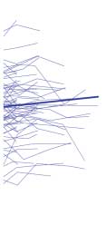
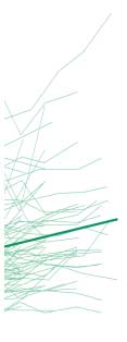
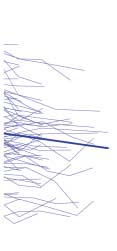
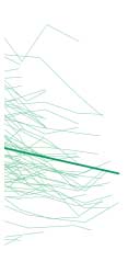
ject brain volume trajectories and treatment tertile group
Longer duration of follow-up was significantly associ-
mean brain volume trajectories for total cerebral WM,
ated with total cerebral tissue, GM, and subcortical brain
lateral ventricles, and frontal GM volumes. We then con-
tissue volume reductions (Table 2; bՅ−0.01 cm3/y), and
ducted an extreme group comparison to contrast brain
with parietal WM, lateral ventricles, and sulcal CSF vol-
volume changes between the most and least treatment
groups. Random regression coefficient mixed model analy-
Because they may be confounders, we adjusted our
ses were duplicated replacing antipsychotic dose with ter-
analysis of treatment effects by statistically correcting for
tile group membership (most vs least treatment) and in-
the effects of follow-up duration, illness severity, and sub-
cluded a tertile groupϫfollow-up duration interaction
stance misuse. Antipsychotic treatment still had signifi-
cant main effects on total cerebral and lobar GM and pu-
For total cerebral WM and lateral ventricles, there were
tamen volumes (Table 2; F Ն 4.33, P Յ .04). Higher
statistically significant main effects of tertile groupϫtime
antipsychotic dose was associated with smaller GM vol-
interaction (Figure, A and B; tՆ2.28, PՅ.02), indicat-
umes (bՅ−0.03) and larger putamen (b=0.01). Anti-
ing that brain volume trajectories differed significantly
psychotic treatment effects on GM volumes were inde-
across tertile groups of antipsychotic treatment. The least-
pendent of follow-up duration (Table 2; antipsychotic
treatment group showed increased total cerebral WM over
treatmentϫfollow-up duration interaction term FՅ1.17,
time in contrast to WM volume reductions among pa-
PՆ.28). On the other hand, there were statistically sig-
tients in the most-treatment group (mean regression
nificant treatmentϫtime interaction effects on total ce-
slopes, 1.30 vs −0.64, respectively). Similarly, patients
rebral tissue volumes, total cerebral and lobar WM, lat-
in the most-treatment group had greater enlargement of
eral ventricles, and sulcal CSF, caudate, putamen, and
lateral ventricles than those in the least-treatment group
cerebellar volumes (Table 2; FՆ3.79, PՅ.05). Higher
(Figure, B; mean regression slopes, 0.39 and 0.16, re-
doses of antipsychotic treatment were associated with
spectively). Consistent with the previous random coef-
greater reductions in WM, caudate, and cerebellar vol-
ficient regression mixed-model analyses where antipsy-
umes over time (bՅ−0.0003), and with greater CSF vol-
chotic treatment was entered as a continuous measure,
ume and putamen enlargements (bՆ0.0008).
extreme tertile group contrast found significant main ef-
To further illustrate how brain volume trajectories may
fects of tertile grouping on frontal GM volumes (t=2.19,
differ according to the amount of antipsychotic treat-
P=.03). Patients who received the most antipsychotic
ment, patients were grouped into tertiles of mean daily
treatment had smaller frontal GM volumes, and this
antipsychotic dose: most treatment (70 patients; mean
difference was independent of follow-up duration
dose, 929.4 CPZ mg equivalents/d), intermediate treat-
(groupϫtime interaction, t=1.32, P=.18).
ment (70 patients; mean dose, 391.7 CPZ mg equivalents/
Patients who received the most antipsychotic treat-
d), and least treatment (71 patients; mean dose, 111.5
ment had smaller baseline total cerebral tissue and larger
CPZ mg equivalents/d). The Figure shows individual sub-
lateral ventricles than the other 2 tertile subgroups
(REPRINTED) ARCH GEN PSYCHIATRY/ VOL 68 (NO. 2), FEB 2011
2011 American Medical Association. All rights reserved. Downloaded From: http://archpsyc.jamanetwork.com/ on 05/24/2013 Most Treatment Intermediate Treatment Least Treatment Most Treatment Intermediate Treatment Least Treatment Most Treatment Intermediate Treatment Least Treatment Figure. Comparison of magnetic resonance imaging brain volume trajectories between tertiles of antipsychotic treatment. Tertiles were categorized as those who
received the most treatment (70 patients; mean [SD] dose, 929.4 [47.7] chlorpromazine [CPZ] mg equivalents/d), intermediate treatment (70 patients; mean [SD]
dose, 391.7 [77.2] CPZ mg equivalents/d), and the least treatment (71 patients; mean [SD] dose, 111.5 [87.7] CPZ mg equivalents/d). Individual patient brain
volume trajectories (thin lines) and treatment tertile group mean brain volume trajectories (thick lines) are shown for total cerebral white matter (A), lateral
ventricles (B), and frontal gray matter volumes (C).
(REPRINTED) ARCH GEN PSYCHIATRY/ VOL 68 (NO. 2), FEB 2011
2011 American Medical Association. All rights reserved. Downloaded From: http://archpsyc.jamanetwork.com/ on 05/24/2013 Table 3. Random Regression Coefficient Mixed Models: Fixed Effects of Typical APSs, Nonclozapine Atypical APSs, and Clozapine on MRI Brain Volumes in 211 Schizophrenia Patientsa Typical APSsb Nonclozapine Atypicalsc Clozapined Regions of Interest
Abbreviations: APSs, antipsychotics; CSF, cerebrospinal fluid; GM, gray matter; MRI, magnetic resonance imaging; WM, white matter.
a Covariates: intracranial volume at intake scan, sex, imaging protocol, age at intake scan, follow-up duration, illness severity (mean Global Assessment Scale
score), and substance misuse; random effects: follow-up duration and an intercept term to model within-patient correlations in brain volumes across time
(unstructured covariance structure).
b Lifetime typical antipsychotic treatment up to the time of MRI scan (mean daily APS treatment; chlorpromazine milligram equivalents per day).
c Lifetime nonclozapine atypical APS treatment up to the time of MRI scan (mean daily APS treatment; chlorpromazine milligram equivalents per day).
d Lifetime clozapine treatment up to the time of MRI scan (mean daily APS treatment; chlorpromazine milligram equivalents per day).
e Estimate of regression coefficient or slope.
(FՆ5.30, PՅ.006). There were no statistically signifi-
repeated the mixed-models analyses in Table 2 by re-
cant differences between the treatment tertile groups re-
placing mean daily (total) antipsychotic dose with life-
garding the other baseline brain volumes (F Յ 2.95,
time mean daily doses of typical antipsychotics, non-
clozapine atypical antipsychotics, and clozapine up untilthe time of each MRI scan (covariates: initial intracra-
INDEPENDENT EFFECTS OF ILLNESS SEVERITY
nial volume, sex, imaging protocol, and age at initial scan;
AND SUBSTANCE ABUSE ON BRAIN VOLUMES
other fixed effects: follow-up duration, mean GAS score,and substance misuse severity; and random effects: fol-
After controlling for the other 3 predictors, mean GAS
low-up duration and intercept term).
score had significant main effects on total cerebral GM
There were significant main effects of typical anti-
and frontal GM volumes (Table 2) (FՆ4.38, PՅ.04). Less
psychotic dose, nonclozapine atypical antipsychotic
illness severity was associated with increased brain tis-
dose, and clozapine dose on GM brain volumes
sue volumes (bՆ0.27). There were no statistically sig-
(Table 3). Higher typical antipsychotic doses were
nificant main effects of mean GAS score on the other brain
associated with smaller total cerebral GM and frontal
volumes (FՅ3.65, PՆ.06).
The majority of the sample (68.3%) did not meet
GM volumes (FՆ4.82, PՅ.03). Higher doses of non-
criteria for alcohol abuse/dependence or illicit drug
clozapine atypical antipsychotics were associated with
abuse/dependence. Seven patients had alcohol abuse/
lower frontal and parietal GM volumes (F Ն 6.74,
dependence only, 13 marijuana abuse/dependence only,
P = .01), and higher clozapine doses were associated
8 alcohol and marijuana abuse/dependence only, 19 al-
with smaller total cerebral and lobar GM volumes
cohol abuse/dependence and nonmarijuana illicit drug
(F Ն 10.90, P Յ .001). For WM volumes, higher non-
abuse/dependence, and 30 nonmarijuana illicit drug abuse/
clozapine atypical antipsychotic doses were significantly
dependence only. Severity of alcohol/illicit substance mis-
associated with larger parietal WM volumes (F=4.34,
use had no significant main effects on brain volumes
P = .04). There were no statistically significant main
(Table 2) (FՅ1.69, PՆ.20) except on lateral ventricles
effects of typical antipsychotic class or nonclozapine
(F = 5.60, P = .02; b = 2.44) and on cerebellar volumes
atypical antipsychotic class on the remaining WM brain
(F=5.48, P=.02; b=−3.25).
volume measures or on lateral ventricles. Higher cloza-pine doses were associated with larger sulcal CSF vol-
INDEPENDENT EFFECTS OF ANTIPSYCHOTIC
umes and smaller caudate, putamen, and thalamic vol-
CLASS ON BRAIN VOLUMES
umes (F Ն 4.70, P Յ .03). Enlarged putamen wasassociated with higher doses of both typical and non-
To explore whether typical antipsychotics, noncloza-
clozapine atypical antipsychotics. Treatment with
pine atypical antipsychotics, and clozapine may have dif-
higher doses of nonclozapine atypical antipsychotics
ferential effects on brain volumes in schizophrenia, we
was also associated with caudate volume enlargement.
(REPRINTED) ARCH GEN PSYCHIATRY/ VOL 68 (NO. 2), FEB 2011
2011 American Medical Association. All rights reserved. Downloaded From: http://archpsyc.jamanetwork.com/ on 05/24/2013
inference, cellular metabolism. Previous positron emis-sion tomography studies conducted by our group52-54confirm that both typical and atypical antipsychotics in-
In this large longitudinal cohort of patients with schizo-
crease putamen cerebral blood flow. In addition, anti-
phrenia (211 patients with 674 MRI scans) who were in
psychotics reduce frontal cerebral blood flow, suggest-
their first episode and had received minimal treatment
ing that chronic frontal hypoperfusion could be a
at the time of entry into the study, we examined the in-
mechanism underlying smaller brain tissue volumes. How-
dependent effects of 4 variables on progressive brain
ever, the available studies that have used morphometric
change during an extended period: illness duration, an-
MRI to examine the effects of antipsychotics on cortical
tipsychotic treatment, illness severity, and substance mis-
GM have yielded ambiguous results,10,55-57 possibly due
use. We found that longer follow-up was associated with
to small sample sizes, differing duration of treatment as-
a greater decrease in brain tissue volumes. Antipsy-
sessment, variation in brain regions measured, and dis-
chotic treatment also had a significant influence on brain
volumes even after accounting for the potential con-
In the present study, WM but not GM volumes showed
founds of the other 3 variables. More antipsychotic treat-
significant timeϫantipsychotic treatment interaction ef-
ment was associated both with generalized tissue vol-
fects. Although higher antipsychotic doses were associ-
ume reduction involving multiple subregions and with
ated with a decrease in GM volumes, this relationship
a specific increase in putamen. The other 2 variables, se-
did not appear to change during the course of longitu-
verity of illness and substance abuse, had minimal or no
dinal follow-up in our study. A change in WM volume
effects. Progressive brain volume changes during the life-
trajectories, on the other hand, was associated with an-
long course of schizophrenia, including GM and WM vol-
tipsychotic treatment. As illustrated by the extreme ter-
ume reductions, CSF volume expansions, and basal gan-
tile treatment group comparisons, patients in the most-
glia volume enlargements, appeared in part to be related
treatment group had longitudinal WM volume reductions.
to antipsychotics. These findings may potentially have
As a group, patients with the least treatment showed WM
clinical implications for the use of long-term antipsy-
volume increases over time that would be expected of
individuals during their third and fourth decades of life.58
The plausibility of long-term antipsychotic treatment
Bartzokis and colleagues59 previously found that pa-
leading to global brain volume reductions is further sup-
tients with schizophrenia do not show the normal age-
ported by recent controlled studies in macaque mon-
related WM volume expansion during early to mid-
keys.19-21 Animal studies provide an additional perspec-
adulthood. Thus, the treatmentϫtime interaction effects
tive on possible causative links because they permit
in the present study suggest that WM volume deficits in
postmortem neuropathological examination of the brain.
schizophrenia may, in part, be related to antipsychotic
Dorph-Petersen et al19 administered haloperidol, olanza-
treatment. Our study found that all 3 classes of antipsy-
pine, or sham medication to macaque monkeys in doses
chotics (ie, typical, nonclozapine atypical, and cloza-
that produced plasma levels equivalent to those observed
pine) were associated with deceases in GM brain vol-
in treatment of schizophrenia patients. After 17 to 27 months
umes. The only differential effects on brain volumes
of treatment, both haloperidol- and olanzapine-treated mon-
between typical and nonclozapine atypical antipsychot-
keys had an equivalent and highly significant 8% to 11%
ics were in parietal WM and caudate measures. These typi-
decrease in fresh brain weight and volume when com-
cal-atypical differential effects on brain volumes in our
pared with the sham group. These decreases affected all ma-
study differ somewhat from a recent randomized treat-
jor brain regions but were most robust in frontal and pa-
ment study.10 Lieberman and colleagues found that halo-
rietal lobes. The neuropathological manifestations of
peridol treatment was associated with progressive GM
antipsychotic-related frontoparietal volume reductions in
volume reductions during that 2-year study. In con-
macaque monkeys involves decreased astrocyte numbers,
trast, olanzapine-treated patients did not show GM vol-
decreased dendritic arborization, decreased dendritic spine
ume decrement. This raises concerns regarding the pos-
density, and increased neuronal density with no neuronal
sibility of typical antipsychotic-associated neurotoxic
loss.20,21 Although there have been some conflicting find-
effects. Our study suggests that atypical antipsychotic
ings in the nonhuman primate literature (eg, studies by
treatment may mitigate parietal WM volume loss in pa-
Lidow et al41 and Sweet et al42), these neuropathological
tients with schizophrenia; this finding needs to be inter-
changes are strikingly similar to those described in the
preted with caution and will require support from addi-
schizophrenia postmortem literature (eg, studies by Pak-
tional well-designed clinical trials in first-episode patients.
Our results must be interpreted in the context of ad-
These findings are also consistent with previous MRI
ditional limitations. Identifying an association does not
studies suggesting that antipsychotics produce changes
necessarily indicate a causal relationship. Furthermore,
in the human brain that are measurable by in vivo neu-
observational studies involving long durations such as
roimaging techniques. The earliest work with morpho-
ours inevitably preclude use of the “gold standard”: a ran-
metric MRI found increased basal ganglia size in schizo-
dom-assignment controlled trial. The current study could
phrenia patients, typically in the putamen.45,46 Subsequent
have been strengthened by having control groups, eg,
studies have shown that this may be a medication effect
schizophrenia patients assigned to deferred or no anti-
and that typical antipsychotics in particular play a causal
psychotic treatment or healthy volunteers treated with
role in basal ganglia enlargement.47-51 Positron emission
antipsychotics for comparable periods. However, ethi-
tomography studies measure cerebral blood flow and, by
cal standards in human subject research prohibit such
(REPRINTED) ARCH GEN PSYCHIATRY/ VOL 68 (NO. 2), FEB 2011
2011 American Medical Association. All rights reserved. Downloaded From: http://archpsyc.jamanetwork.com/ on 05/24/2013
comparison groups. The small number of schizophre-
cluding children, the elderly, and patients with bipolar dis-
nia patients in our sample who received no antipsy-
order or depressive disorders.27-30 They are also used in ado-
chotic treatment did not allow for meaningful statistical
lescents who have been identified to be at high risk for
analyses. Illness severity and antipsychotic dosages were
schizophrenia. Our findings may lead to heightened con-
modestly correlated (Spearman r=−0.21), and patients
cerns regarding potential brain volume changes associ-
who received the most treatment had smaller baseline ce-
ated with the sharp rise in atypical antipsychotic use in non-
rebral tissue volumes. Associations between smaller brain
schizophrenia psychiatric disorders. Even though no studies
tissue volumes and more antipsychotic treatment may still
have assessed the long-term effects of antipsychotics on brain
be moderated via illness severity despite our inclusion
volumes in nonschizophrenia patients, our results sug-
of illness severity as a covariate and obtaining similar re-
gest that antipsychotics should still be used with caution
sults from different measures of illness severity. Even with
in these patient groups after careful risk-benefit assess-
the most sophisticated statistical methods, we may not
ment. Because typical antipsychotics are off patent and less
be able to fully distinguish the potential confounding in-
expensive than atypical ones, there is also a growing trend
fluences that illness severity or other sources of unmea-
to prescribe them preferentially for patients with schizo-
sured variance could still have on the relationships
phrenia. Given that these older medications carry a greater
between progressive brain volume reductions and anti-
risk of producing extrapyramidal adverse effects and tar-
psychotic treatment. Last, although our Talairach atlas–
dive dyskinesia, such a shift in clinical practice may pro-
based lobar measures have well-established reliability and
duce deleterious effects on the primary diseased organ in
validity, these cerebral brain volumes lack precision to
delineate abnormalities within smaller subregions im-plicated in schizophrenia (eg, superior temporal gyrus
Findings from the present study raise several clinical
Antipsychotics are effective medications for reducing some
questions. Are antipsychotic-associated GM and WM vol-
of the target clinical symptoms of schizophrenia: psy-
ume reductions “bad” for patients? The implicit assump-
chotic symptoms. In medicine we are aware of many in-
tion is that brain volume reductions are probably unde-
stances in which improving target symptoms worsens
sirable because patients with schizophrenia already have
other symptoms. Hormone therapy relieves meno-
diffuse brain volume deficits at the time of illness onset.
pausal symptoms but increases stroke risk. Nonsteroi-
Schizophrenia patients with poor outcomes are also more
dal anti-inflammatory drugs relieve pain but increase the
likely to have smaller brain volumes. However, the neu-
likelihood of duodenal ulcers and gastrointestinal tract
robiological changes that underlie MRI measurements of
bleeding. It is possible that, although antipsychotics re-
antipsychotic-associated brain volume decrement re-
lieve psychosis and its attendant suffering, these drugs
main poorly understood. Some studies indicate that an-
may not arrest the pathophysiologic processes underly-
tipsychotic-induced changes mimic the neuropathologi-
ing schizophrenia and may even aggravate progressive
cal changes of schizophrenia,20,21 while others suggest
otherwise.41,42 If antipsychotics do indeed result in del-eterious brain tissue volume reductions, how does thisinfluence the risk-benefit ratio of antipsychotic treat-
Submitted for Publication: March 5, 2010; final revi-
ment? Given that these medications have substantially
sion received September 14, 2010; accepted September
improved the long-term prognosis of schizophrenia and
that schizophrenia is a disease with significant morbid-
Correspondence: Beng-Choon Ho, MRCPsych, Depart-
ity, continued use of antipsychotics is clearly still nec-
ment of Psychiatry, W278 GH, University of Iowa Carver
essary. However, our findings point toward the impor-
College of Medicine, 200 Hawkins Dr, Iowa City, IA
tance of prescribing the lowest doses necessary to control
symptoms. They also imply the need for rethinking the
Author Contributions: Drs Ho and Andreasen had full
underlying pathological processes in schizophrenia,47,48
access to all the data in the study and take responsibility
the target at which treatment is aimed, and the possibil-
for the integrity of the data and the accuracy of the data
ity that antipsychotic treatment may improve psychotic
symptoms but also contribute to progressive brain tis-
Financial Disclosure: Dr Ho receives grant support from
sue volume deficits. Antipsychotics were designed for the
Ortho-McNeil Janssen Scientific Affairs. Dr Andreasen
purpose indicated by their name, ie, to arrest psychosis.
has served on the Ortho-McNeil Janssen Advisory Board
Not only is it probable that antipsychotics do not treat
and receives grant support from Ortho-McNeil Janssen
the fundamental pathophysiologic mechanism of schizo-
phrenia (ie, the brain disease), but we perhaps must also
Funding/Support: This research was supported in part
entertain the possibility that they might have poten-
by grants MH68380, MH31593, MH40856, and MH43271
tially undesirable effects of brain tissue volume reduc-
from the National Institute of Mental Health.
tions. In conjunction with neuroscientists and clinical
Role of the Sponsor: The National Institute of Mental
investigators, pharmaceutical companies must con-
Health financially supported the design and conduct of
tinue the vigorous search for agents that are genuinely
the study; the collection, management, analysis, and in-
terpretation of the data; and the preparation of the manu-
The second-generation antipsychotics are also now
script, but was not involved in manuscript review or
widely used for people who do not have schizophrenia, in-
(REPRINTED) ARCH GEN PSYCHIATRY/ VOL 68 (NO. 2), FEB 2011
2011 American Medical Association. All rights reserved. Downloaded From: http://archpsyc.jamanetwork.com/ on 05/24/2013 Online-Only Material: The supplementary Methods and
fluence of chronic exposure to antipsychotic medications on brain size before
References and the eFigure and eTables are available at
and after tissue fixation: a comparison of haloperidol and olanzapine in ma-
caque monkeys. Neuropsychopharmacology. 2005;30(9):1649-1661.
20. Konopaske GT, Dorph-Petersen KA, Pierri JN, Wu Q, Sampson AR, Lewis DA. Additional Contributions: Dawei Liu, PhD, provided
Effect of chronic exposure to antipsychotic medication on cell numbers in the
valuable advice and assistance in statistical analysis.
parietal cortex of macaque monkeys. Neuropsychopharmacology. 2007;32(6):1216-1223.
21. Konopaske GT, Dorph-Petersen K-A, Sweet RA, Pierri JN, Zhang W, Sampson
AR, Lewis DA. Effect of chronic antipsychotic exposure on astrocyte and oligo-dendrocyte numbers in macaque monkeys. Biol Psychiatry. 2008;63(8):759-765.
1. Murray CJL, Lopez AD, eds. The Global Burden of Disease: A Comprehensive
22. Navari S, Dazzan P. Do antipsychotic drugs affect brain structure? a systematic
Assessment of Mortality and Disability From Diseases, Injuries, and Risk Fac-
and critical review of MRI findings. Psychol Med. 2009;39(11):1763-1777. tors in 1990 and Projected to 2020. Cambridge, MA: Harvard University Press;
23. Smieskova R, Fusar-Poli P, Allen P, Bendfeldt K, Stieglitz RD, Drewe J, Radue
EW, McGuire PK, Riecher-Rössler A, Borgwardt SJ. The effects of antipsychot-
2. Gilbert PL, Harris MJ, McAdams LA, Jeste DV. Neuroleptic withdrawal in schizo-
ics on the brain: what have we learnt from structural imaging of schizophrenia?
phrenic patients: a review of the literature. Arch Gen Psychiatry. 1995;52(3):
a systematic review. Curr Pharm Des. 2009;15(22):2535-2549.
24. Borgwardt SJ, Smieskova R, Fusar-Poli P, Bendfeldt K, Riecher-Rössler A.
3. DeLisi LE, Sakuma M, Tew W, Kushner M, Hoff AL, Grimson R. Schizophrenia
The effects of antipsychotics on brain structure: what have we learnt from struc-
as a chronic active brain process: a study of progressive brain structural change
tural imaging of schizophrenia? Psychol Med. 2009;39(11):1781-1782.
subsequent to the onset of schizophrenia. Psychiatry Res. 1997;74(3):129-
25. Lewis DA. Brain volume changes in schizophrenia: how do they arise? what do
they mean? Psychol Med. 2009;39(11):1779-1780.
4. Gur RE, Cowell P, Turetsky BI, Gallacher F, Cannon T, Bilker W, Gur RC. A fol-
26. Vita A, De Peri L. The effects of antipsychotic treatment on cerebral structure
low-up magnetic resonance imaging study of schizophrenia: relationship of neu-
and function in schizophrenia. Int Rev Psychiatry. 2007;19(4):429-436.
roanatomical changes to clinical and neurobehavioral measures. Arch Gen
27. Domino ME, Swartz MS. Who are the new users of antipsychotic medications?
Psychiatry. 1998;55(2):145-152. Psychiatr Serv. 2008;59(5):507-514.
5. Lieberman J, Chakos M, Wu H, Alvir J, Hoffman E, Robinson D, Bilder R. Lon-
gitudinal study of brain morphology in first episode schizophrenia. Biol Psychiatry.
28. Olfson M, Blanco C, Liu L, Moreno C, Laje G. National trends in the outpatient
treatment of children and adolescents with antipsychotic drugs. Arch Gen
6. Mathalon DH, Sullivan EV, Lim KO, Pfefferbaum A. Progressive brain volume
Psychiatry. 2006;63(6):679-685.
changes and the clinical course of schizophrenia in men: a longitudinal mag-
29. Castle NG, Hanlon JT, Handler SM. Results of a longitudinal analysis of national
netic resonance imaging study. Arch Gen Psychiatry. 2001;58(2):148-157.
data to examine relationships between organizational and market characteris-
7. Cahn W, Hulshoff Pol HE, Lems EB, van Haren NE, Schnack HG, van der Linden
tics and changes in antipsychotic prescribing in US nursing homes from 1996
JA, Schothorst PF, van Engeland H, Kahn RS. Brain volume changes in first-
through 2006. Am J Geriatr Pharmacother. 2009;7(3):143-150.
episode schizophrenia: a 1-year follow-up study. Arch Gen Psychiatry. 2002;
30. Governale L, Mehta H. Outpatient use of atypical antipsychotic agents in the pe-
diatric population: years 2004-2008. US Food and Drug Administration Web site.
8. Ho BC, Andreasen NC, Nopoulos P, Arndt S, Magnotta V, Flaum M. Progressive
http://www.fda.gov/downloads/AdvisoryCommittees/CommitteesMeetingMaterials
structural brain abnormalities and their relationship to clinical outcome: a lon-
/PediatricAdvisoryCommittee/UCM193204.pdf. Published December 8, 2009. Ac-
gitudinal magnetic resonance imaging study early in schizophrenia. Arch GenPsychiatry. 2003;60(6):585-594.
31. Flaum MA, Andreasen NC, Arndt S. The Iowa prospective longitudinal study of
9. Kasai K, Shenton ME, Salisbury DF, Hirayasu Y, Onitsuka T, Spencer MH, Yurgelun-
recent-onset psychoses. Schizophr Bull. 1992;18(3):481-490.
Todd DA, Kikinis R, Jolesz FA, McCarley RW. Progressive decrease of left Heschl
32. Andreasen NC. The Scale for Assessment of Negative Symptoms (SANS). Iowa
gyrus and planum temporale gray matter volume in first-episode schizophrenia:
a longitudinal magnetic resonance imaging study. Arch Gen Psychiatry. 2003;
33. Andreasen NC. The Scale for Assessment of Positive Symptoms (SAPS). Iowa
10. Lieberman JA, Tollefson GD, Charles C, Zipursky R, Sharma T, Kahn RS, Keefe
34. Andreasen NC, Flaum M, Arndt S. The Comprehensive Assessment of Symp-
RS, Green AI, Gur RE, McEvoy J, Perkins D, Hamer RM, Gu H, Tohen M; HGDH
toms and History (CASH): an instrument for assessing diagnosis and
Study Group. Antipsychotic drug effects on brain morphology in first-episode
psychopathology. Arch Gen Psychiatry. 1992;49(8):615-623.
psychosis. Arch Gen Psychiatry. 2005;62(4):361-370.
35. Andreasen NC. PSYCH-BASE. Iowa City: University of Iowa; 1989.
11. Kasai K, Shenton ME, Salisbury DF, Hirayasu Y, Lee CU, Ciszewski AA, Yurgelun-
36. Ho BC, Flaum M, Hubbard W, Arndt S, Andreasen NC. Validity of symptom as-
Todd D, Kikinis R, Jolesz FA, McCarley RW. Progressive decrease of left supe-
sessment in psychotic disorders: information variance across different sources
rior temporal gyrus gray matter volume in patients with first-episode schizophrenia.
of history. Schizophr Res. 2004;68(2-3):299-307. Am J Psychiatry. 2003;160(1):156-164.
37. Andreasen NC, Nopoulos P, Magnotta V, Pierson R, Ziebell S, Ho BC. Progres-
12. van Haren NE, Hulshoff Pol HE, Schnack HG, Cahn W, Mandl RC, Collins DL,
sive neural change in schizophrenia: a prospective longitudinal study of first epi-
Evans AC, Kahn RS. Focal gray matter changes in schizophrenia across the course
sode schizophrenia. Biol Psychiatry. In press.
of the illness: a 5-year follow-up study. Neuropsychopharmacology. 2007;
38. Davis JM. Dose equivalence of the antipsychotic drugs. J Psychiatr Res. 1974;
13. Cahn W, Rais M, Stigter FP, van Haren NE, Caspers E, Hulshoff Pol HE, Xu Z,
39. Andreasen NC, Pressler M, Nopoulos P, Miller D, Ho BC. Antipsychotic dose equiva-
Schnack HG, Kahn RS. Psychosis and brain volume changes during the first five
lents and dose-years: a standardized method for comparing exposure to differ-
years of schizophrenia. Eur Neuropsychopharmacol. 2009;19(2):147-151.
ent drugs. Biol Psychiatry. 2010;67(3):255-262.
14. Mathalon DH, Rapoport JL, Davis KL, Krystal JH. Neurotoxicity, neuroplasticity,
40. Endicott J, Spitzer RL, Fleiss JL, Cohen J. The Global Assessment Scale: a pro-
and magnetic resonance imaging morphometry [letter]. Arch Gen Psychiatry. 2003;
cedure for measuring overall severity of psychiatric disturbance. Arch GenPsychiatry. 1976;33(6):766-771.
15. Weinberger DR, McClure RK. Neurotoxicity, neuroplasticity, and magnetic reso-
41. Lidow MS, Song Z-M, Castner SA, Allen PB, Greengard P, Goldman-Rakic PS.
nance imaging morphometry [reply]. Arch Gen Psychiatry. 2003;60(8):848-849.
Antipsychotic treatment induces alterations in dendrite- and spine-associated pro-
16. Hulshoff Pol HE, Kahn RS. What happens after the first episode? a review of pro-
teins in dopamine-rich areas of the primate cerebral cortex. Biol Psychiatry. 2001;
gressive brain changes in chronically ill patients with schizophrenia. Schizophr
42. Sweet RA, Henteleff RA, Zhang W, Sampson AR, Lewis DA. Reduced dendritic spine
17. Pantelis C, Yücel M, Wood SJ, Velakoulis D, Sun D, Berger G, Stuart GW, Yung
density in auditory cortex of subjects with schizophrenia. Neuropsychopharmacology.
A, Phillips L, McGorry PD. Structural brain imaging evidence for multiple patho-
logical processes at different stages of brain development in schizophrenia.
43. Pakkenberg B. Total nerve cell number in neocortex in chronic schizophrenics
Schizophr Bull. 2005;31(3):672-696.
and controls estimated using optical disectors. Biol Psychiatry. 1993;34(11):
18. Ho BC, Andreasen NC, Dawson JD, Wassink TH. Association between brain-
derived neurotrophic factor Val66Met gene polymorphism and progressive brain
44. Selemon LD, Rajkowska G, Goldman-Rakic PS. Abnormally high neuronal den-
volume changes in schizophrenia. Am J Psychiatry. 2007;164(12):1890-1899.
sity in the schizophrenic cortex: a morphometric analysis of prefrontal area 9
19. Dorph-Petersen KA, Pierri JN, Perel JM, Sun Z, Sampson AR, Lewis DA. The in-
and occipital area 17. Arch Gen Psychiatry. 1995;52(10):805-818.
(REPRINTED) ARCH GEN PSYCHIATRY/ VOL 68 (NO. 2), FEB 2011
2011 American Medical Association. All rights reserved. Downloaded From: http://archpsyc.jamanetwork.com/ on 05/24/2013
45. Jernigan TL, Zisook S, Heaton RK, Moranville JT, Hesselink JR, Braff DL. Mag-
53. Miller DD, Andreasen NC, O’Leary DS, Rezai K, Watkins GL, Ponto LL, Hichwa
netic resonance imaging abnormalities in lenticular nuclei and cerebral cortex in
RD. Effect of antipsychotics on regional cerebral blood flow measured with posi-
schizophrenia. Arch Gen Psychiatry. 1991;48(10):881-890.
tron emission tomography. Neuropsychopharmacology. 1997;17(4):230-
46. Swayze VW II, Andreasen NC, Alliger RJ, Yuh WT, Ehrhardt JC. Subcortical and
temporal structures in affective disorder and schizophrenia: a magnetic reso-
54. Corson PW, O’Leary DS, Miller DD, Andreasen NC. The effects of neuroleptic medi-
nance imaging study. Biol Psychiatry. 1992;31(3):221-240.
cations on basal ganglia blood flow in schizophreniform disorders: a compari-
47. Chakos MH, Lieberman JA, Alvir J, Bilder R, Ashtari M. Caudate nuclei volumes
son between the neuroleptic-naïve and medicated states. Biol Psychiatry. 2002;
in schizophrenic patients treated with typical antipsychotics or clozapine. Lancet.
55. Crespo-Facorro B, Kim J-J, Chemerinski E, Magnotta V, Andreasen NC, Nopou-
48. Corson PW, Nopoulos P, Miller DD, Arndt S, Andreasen NC. Change in basal gan-
los P. Morphometry of the superior temporal plane in schizophrenia: relation-
glia volume over 2 years in patients with schizophrenia: typical versus atypical
ship to clinical correlates. J Neuropsychiatry Clin Neurosci. 2004;16(3):284-
neuroleptics. Am J Psychiatry. 1999;156(8):1200-1204.
49. Frazier JA, Giedd JN, Kaysen D, Albus K, Hamburger S, Alaghband-Rad J, Lenane
MC, McKenna K, Breier A, Rapoport JL. Childhood-onset schizophrenia: brain
56. McCormick L, Decker L, Nopoulos P, Ho BC, Andreasen N. Effects of atypical
MRI rescan after 2 years of clozapine maintenance treatment. Am J Psychiatry.
and typical neuroleptics on anterior cingulate volume in schizophrenia. Schizophr
50. Gur RE, Maany V, Mozley PD, Swanson C, Bilker W, Gur RC. Subcortical MRI
57. Pressler M, Nopoulos P, Ho BC, Andreasen NC. Insular cortex abnormalities in
volumes in neuroleptic-naive and treated patients with schizophrenia. Am J
schizophrenia: relationship to symptoms and typical neuroleptic exposure. BiolPsychiatry. 1998;155(12):1711-1717. Psychiatry. 2005;57(4):394-398.
51. Keshavan MS, Rosenberg D, Sweeney JA, Pettegrew JW. Decreased caudate vol-
58. Sowell ER, Peterson BS, Thompson PM, Welcome SE, Henkenius AL, Toga AW.
ume in neuroleptic-naive psychotic patients. Am J Psychiatry. 1998;155(6):
Mapping cortical change across the human life span. Nat Neurosci. 2003;6
52. Miller DD, Andreasen NC, O’Leary DS, Watkins GL, Boles Ponto LL, Hichwa RD.
59. Bartzokis G, Nuechterlein KH, Lu PH, Gitlin M, Rogers S, Mintz J. Dysregulated
Comparison of the effects of risperidone and haloperidol on regional cerebral
brain development in adult men with schizophrenia: a magnetic resonance imaging
blood flow in schizophrenia. Biol Psychiatry. 2001;49(8):704-715.
study. Biol Psychiatry. 2003;53(5):412-421.
(REPRINTED) ARCH GEN PSYCHIATRY/ VOL 68 (NO. 2), FEB 2011
2011 American Medical Association. All rights reserved. Downloaded From: http://archpsyc.jamanetwork.com/ on 05/24/2013
Gender Differences in Sleep, Fatigue, and Daytime Activity ina Pediatric Oncology Sample Receiving DexamethasoneStacy D. Sanford, PHD, James O. Okuma, MS, Jianmin Pan, PHD, Deo Kumar Srivastava, PHD,Nancy West, BSN, Lynne Farr, PHD and Pamela S. Hinds, PHD, FAANSt Jude Children’s Research Hospital, MemphisObjective To examine gender differences in sleep, fatigue, and daytime activity in a samp
Bericht über den "13. Internationalen Kongress der Movement Disorder Socie- ty" in Paris Vom 7. bis 11. Juni 2009 trafen sich Ärzte und Wissenschaftler aus der ganzen Welt zum"13. Internationalen Kongress der Movement Disorder Society" in Paris, um über die neues-ten Forschungsergebnisse aus dem Bereich der Bewegungsstörungen zu diskutieren. ImRahmen des fünftägigen Ko


 Most Treatment