Home/my story/Patient stories/Error database/Hospital wards/Have your say/Library
Venous Sinus Thrombosis Explanation of medical Terminology
Venous: deoxygenated blood, returning to the heartSinus: cavity, space, gapThrombosis: blood clot
What is the normal functioning of this organ?
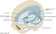
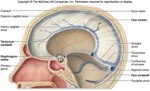
The dura mater is the outermost, of the three layers of skin (meninges) surrounding the brain and spinal cord. The two other layers in are the arachnoid mater (spider like layer of skin) and the pia mater (close hugging layer of skin). The dura mater is actually a double sided layer of skin. It’s outer layer is attached to the inside of the skull. It’s inner layer forms a tent that surrounds and protects the soft brain. There are areas in the brain where the two layers of the dura mater separate out, creating spaces or sinuses.
The deoxygenated (venous) blood flow from the brain, back to the heart is via two channels. The first channel is via these small channels or sinuses in between the two layers of the dura mater. The walls of the dura mata actually form the cavity that carries venous blood away from the capillaries and towards large veins. The second channel is deep veins in the brain (superior cerebral veins, inferior cerebral veins, middle cerebral vein). They carry venous blood away from sinuses, towards the internal jugular veins and back to the heart. What is this disease?
A venous blood clot forms in the dural sinuses. It can be due to blood clotting disorders, trauma or disease. This clot then blocks the normal venous blood flow. The result is a rise in intra cranial pressure. Fluid on the brain (hydrocephalus) can also form. If not treated quickly, death will result. Who get’s it?
Venous sinus thrombosis is a rare condition. It is considered a form of stroke. There are three major causes:
Blood disorders. Diseases that affect the clotting of the blood. Dehydration. Hormone disturbances. Cancer. Traumatic damage Road or other physical trauma. Infections. Surgical procedures to the head and neckInflammatory diseases Lupus, inflammatory bowel disease
Home/my story/Patient stories/Error database/Hospital wards/Have your say/Library
How is it diagnosed?
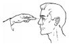
Presentations may include ‘thunderclap’ headache, nausea and vomiting. Other presentations may include abnormal vision, weakness on one side of the face or body. 40% of cases present with seizures. Severe drop in conscious state in cases of raised ICP
CT brain using intravenous dye can be very effective in diagnosing this condition. MRI brain is also used. Cerebral angiography may show up smaller clots.
uTube clip showing scans of venous sinus thrombosis
How is it treated?
Few people realize that there are dramatic differences in the standards of hospitals across Australia. Learn about specialization, privatization and safety standards. (Link to PDF) Which hospital is best for you?
From medication errors to over-treatment to personal bankruptcy, mistakes in the health system are more common that most people realize. Learn how to minimize them. (Link to PDF) Questions to ask your doctor?
It is important that a patient receive prompt medical assessment including neurological examination after any head trauma.
uTube clip of medical assessment of head injured patient coming to emergency department
Herniation due to increased ICP is the major cause of death. Anti coagulation is main therapy. De-compressive surgery can be considered or removal of the clot may be considered. Heparin is commonly used to thin the blood. Heparin can be quickly reversed with the drug protamine if urgent surgery is required.
The anti coagulation (clot breaking) drug heparin is given intravenously, followed by the oral drug warfarin, providing there are no other bleeding risks that would make this treatment unsuitable. Some times anti coagulation is not recommended if there is extensive bleeding as well as the clot. The time spend on warfarin depends on the cause of the clot. Generally if the cause is temporary (such as hormone changes due to pregnancy) the warfarin will be for a short period such as three months. However if the cause is a long term condition (such as a blood clotting disorder) the warfarin may be continued permanently.
Home/my story/Patient stories/Error database/Hospital wards/Have your say/Library
uTube clip showing patient with venous sinus thrombosis being given treatment
It is important that the patient being monitored after a head injury is regularly assessed using a neurological test called the Glasgow Coma Scale.
uTube clip of nurse doing GCS with patient with head injury
Any patient with a brain injury is at risk of brain swelling, causing increased pressure inside the hard skull and tough inflexible dura mater.
uTube clip of raised intracranial pressure
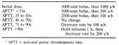
Management of intra venous heparin. Bolus dose 5,000 units IV heparin, then commence heparin infusion based on body weight. APTT should be measured 6 hourly until APPT is within required range. In general APPT is kept between 70 - 90 seconds for full anticoagulation. Changes to this can be made by the doctor.
uTube clip showing heparin dosages and administration
Allied health management
A physiotherapist takes the patient is taken for walks post surgery to reduce risk of pneumonia and to prevent muscle wastage. A physiotherapist will also commence rehabilitation with games and aids to try and restore brain function.
A social worker will discuss financial issues and can organise referrals to CenterLink and possibly the Transport Accident Commission. A social worker can also organise family meetings with the doctors, to help understand the disease and medical treatments.
An occupational therapist can do home assessments to see if it is safe for the patient to return home. This involves looking at thinks like ramps for wheelchairs, rails along walls and equipment to assist with toileting and washing. They can also organise aids to help pick up clothes, put on shoes and manage daily tasks
Discharge planning
Proper discharge planning is proven to be essential to preventing further illness and re admissions to hospital. (Link to PDF) discharge planning
Home/my story/Patient stories/Error database/Hospital wards/Have your say/Library
Making sure you get the best care
In order to make sure your treatment is up to date, have a look at what senior doctors and nurses suggest is the best way to manage your disease(Link) Best Practice Clinical Guidelines - stroke
Make sure your nursing care is up to the standards set by major public hospitals in Australia(Link) Public Hospital policy and procedure manual - Neurosurgery
Statistics
Venous sinus thrombosis is considered a rare form of stroke. It is most common in the third decade. 75% of patients are female. Use of contraceptives by women is suggested as being a cause of the sex imbalance. As this is a rare disorder accurate mortality rates are difficult to obtain, 6 - 16% mortality is suggested, brain herniation is the main cause of death. References
WikipediaCerebral venous sinus thrombosis
National Institute for Health and Care Excellence UKQuick reference guide Stroke July 2008
Resources
NIH Public Access Author Manuscript Discov Med . Author manuscript; available in PMC 2011 November 1. Discov Med. 2010 November ; 10(54): 434–442. Neurorestorative Treatments for Traumatic Brain Injury Ye Xiong 1, Asim Mahmood 1, and Michael Chopp 2,3,* 1 Department of Neurosurgery, Henry Ford Health System, 2799 West Grand Boulevard, Detroit, MI 48202, USA 2 Department of Neurolog

 Home/my story/Patient stories/Error database/Hospital wards/Have your say/Library
Venous Sinus Thrombosis
Home/my story/Patient stories/Error database/Hospital wards/Have your say/Library
Venous Sinus Thrombosis

 Home/my story/Patient stories/Error database/Hospital wards/Have your say/Library
How is it diagnosed?
Home/my story/Patient stories/Error database/Hospital wards/Have your say/Library
How is it diagnosed?
 Home/my story/Patient stories/Error database/Hospital wards/Have your say/Library
uTube clip showing patient with venous sinus thrombosis being given treatment
It is important that the patient being monitored after a head injury is regularly assessed using a neurological test called the Glasgow Coma Scale.
uTube clip of nurse doing GCS with patient with head injury
Any patient with a brain injury is at risk of brain swelling, causing increased pressure inside the hard skull and tough inflexible dura mater.
uTube clip of raised intracranial pressure
Management of intra venous heparin. Bolus dose 5,000 units IV heparin, then commence heparin infusion based on body weight. APTT should be measured 6 hourly until APPT is within required range. In general APPT is kept between 70 - 90 seconds for full anticoagulation. Changes to this can be made by the doctor.
Home/my story/Patient stories/Error database/Hospital wards/Have your say/Library
uTube clip showing patient with venous sinus thrombosis being given treatment
It is important that the patient being monitored after a head injury is regularly assessed using a neurological test called the Glasgow Coma Scale.
uTube clip of nurse doing GCS with patient with head injury
Any patient with a brain injury is at risk of brain swelling, causing increased pressure inside the hard skull and tough inflexible dura mater.
uTube clip of raised intracranial pressure
Management of intra venous heparin. Bolus dose 5,000 units IV heparin, then commence heparin infusion based on body weight. APTT should be measured 6 hourly until APPT is within required range. In general APPT is kept between 70 - 90 seconds for full anticoagulation. Changes to this can be made by the doctor.

 Home/my story/Patient stories/Error database/Hospital wards/Have your say/Library
Making sure you get the best care
Home/my story/Patient stories/Error database/Hospital wards/Have your say/Library
Making sure you get the best care