Asnc imaging guidelines for nuclear cardiology procedures - standardized reporting of radionuclide myocardial perfusion and function
Standardized reporting of radionuclidemyocardial perfusion and function
Peter L. Tilkemeier, MD,a C. David Cooke, MSEE,b Gabriel B. Grossman, MD,PhD,c,d Benjamin D. McCallister Jr, MD,e and R. Parker Ward, MDf
multiple formats consisting of clear and defined struc-tured data elements.
The myocardial perfusion imaging report is the final
The appearance of the standardized report can,
product of a complicated process designed to produce
and should, vary from user to user. Report structure
high quality and valuable patient data. As such, it must
does not rely on a single standard appearance, but
contain sufficient information to convey the details of
rather on the content and utilization of structured data
the procedure while simultaneously remaining succinct.
elements. For example, one laboratory may choose to
The report should provide a basic ‘‘bottom line’’ result
use a report that is formatted in paragraphs while
to the referring physician.-It should follow a defined
another may use a tabular format, or even a combi-
structure and contain standardized data elements so that
nation of the two. All are structured reports as long as
test results are portable as patients move through the
the data are derived from defined, structured data
healthcare system. The structured report is an integral
part of the electronic health care record, and is necessary
Reports have limited utility if they cannot be used
for defining quality in all nuclear cardiology practices.
to communicate and transfer information from one
As the healthcare community moves away from open
source to another. The use of structured data and
reporting, the implementation of structured reporting is
reporting, as defined by the Digital Imaging and
expected to improve quality and outcomes and to reduce
and Integrating the Healthcare Enterprise (IHE) stan-
The myocardial perfusion imaging report is used for
dards, allows this communication to occur. The
various purposes including patient care, research, quality
DICOM standard for stress reporting includes the data
assurance, reimbursement documentation, and incorpo-
elements for structured nuclear cardiology reporting.
ration of data into integrated imaging databases. The
These elements have been adopted by developers and
report may also be used by a variety of interested parties
including physicians, researchers, insurers, and industry.
implementation. IHE has also published a standard for
To accommodate all of these functions and users, one
the communication of data among different vendor
should be able to rearrange the imaging report into
The American College of Cardiology (ACC) has also
published two relevant reporting documents. The‘‘Health Policy Statement on Structured Reporting inCardiovascular Imaging’’defines the generally accepted
From Miriam Hospital,a Providence, RI; Emory University Hospital,b
position of the cardiovascular community regarding
Atlanta, GA; Hospital Moinhos de Vento,c Rio Grande do Sul,Brazil; Cardionuclear-Instituto de Cardiologia,d Porto Alegre, Bra-
structured reporting. The ‘‘Key Data Elements and Def-
zil; Michigan Heart and Vascular Institute,e Ypsilanti, MI;
initions for Cardiac Imaging: A Report of the American
University of Chicago,f Chicago, IL.
College of Cardiology/American Heart Association Task
Unless reaffirmed, retired, or amended by express action of the Board
Force on Clinical Data Standardsis designed to
of Directors of the American Society of Nuclear Cardiology, thisImaging Guideline shall expire as of May 2014.
facilitate the reporting of imaging studies in multi-
Reprint requests: Peter L. Tilkemeier, MD, Miriam Hospital, Provi-
modality environments by coordinating the definitions of
some data elements used for myocardial perfusion and
functional imaging. This has resulted in a ‘‘redefinition’’
Copyright Ó 2009 by the American Society of Nuclear Cardiology.
of some of the data elements in prior versions of
ASNC imaging guidelines for nuclear cardiology procedures
the nuclear cardiology standard. This updated image
reporting guideline incorporates and harmonizes these
recommendations, unifies prior ASNC documents, andexpands the imaging guidelines to include exercise and
resting first-pass radionuclide angiography (FPRNA),
equilibrium radionuclide angiocardiography (ERNA),
positron emission tomography (PET), and viability
This document will address both structured reports
as well as structured data. Structured data elements will
be reported in tables pertaining to each of the broad
areas a structured report should contain. These tables
consist of the variables, their description, format (i.e.,
text, numeric, date), priority (i.e., required, recom-
mended, or optional), and allowed response(s). The
structured reports portion of the document will discuss
the use of the standard data elements to construct a
Due to considerable recent development and matur-
ing of the tools required for widespread clinical
implementation of structured reporting, the current doc-
ument is an update of an earlier image reporting guideline
that was developed by the American Society of Nuclear
Cardiology (ASNC).Given this significant degree of
development, ASNC supports the mandatory use ofstructured reporting using standardized data elements in
ECG, Electrocardiographic; LV, left ventricular; RV, right
myocardial perfusion imaging reports. This should be
ventricular; FPRNA, first-pass radionuclide angiography;
implemented as part of the laboratory accreditation
ERNA, equilibrium radionuclide angiocardiography.
This publication is designed to provide imaging
guidelines for physicians and technologists who are
A few of the general data elements, and many of the
qualified to practice nuclear cardiology. While the
specific data elements, may be recorded at the time that
information supplied in this document has been care-
the test is performed. Some elements may not be
fully reviewed by experts in the field, the document
required in the final report. This may be the case for
should not be considered medical advice or a profes-
some fields that are required for quality reporting, but
not necessarily for reporting the findings from an indi-vidual patient’s study for specific patient management.
Many different structured reports can be generated
from a set of structured data. The potential reports include:a clinical patient-specific report, summary quality
report, billing report, and other reports as needed. The
According to the ‘‘Health Policy Statement on
greatest strength to structured data utilization is the ability
Structured Reporting in Cardiovascular Imaging,’’the
to generate multiple report formats with varying levels of
standard components of a report include the following
detail depending on the clinical or administrative need.
major headings: Administrative Information, Patient
This document will harmonize these generalized
Demographics, Study Referral Data, History and Risk
concepts and apply them specifically to nuclear cardi-
Factors, Study Description, Study Findings, and other
ology. Due to the variability of the study types
reporting parameters. These elements are outlined in
encompassed by this document, some of the data ele-
detail in ‘‘Key Data Elements and Definitions for Car-
ments are specific to certain types of acquisitions, or are
diac Imaging: A Report of the American College of
dependent upon the study indication (e.g., viability
Cardiology/American Heart Association Task Force on
determination by PET imaging). Therefore, some data
Clinical Data Standardswhich addresses specific
elements may be required for certain acquisitions and
details for each of these major headings for multiple
clinical indications, while some may be optional and
perhaps irrelevant for other indications.
ASNC imaging guidelines for nuclear cardiology procedures
A number of the data elements contained in the
tables have been derived from, and harmonized with,
The Patient Demographics and Study Referral data
other guideline documents, some multi-societal and
section provides the clinical indications for the study.
others ASNC-specific.This update also addresses
Major areas to be considered are: diagnosis of coronary
additional modalities that were not included in the
artery disease (CAD), extent and severity of known CAD,
prior versions of the document, such as: PET, viabil-
risk stratification, determination of viability, and assess-
ity, FPRNA, and ERNA both at rest and with
ment of acute chest pain syndromes. With the inclusion of
exercise. The data elements required for reporting the
the History and Risk Factors section, this would complete
additional modalities have been added to specific
the data elements contained in Tables and .
tables where appropriate or additional tables have
The specific purpose for which the test is being per-
been added to the document to cover those items that
formed must be clearly identified. This provides the
were specific to the modality and could not be
required documentation for the medical necessity of the
generalized to one of the existing table headings
study and its appropriateness, and focuses the report on
the question asked by the referring physician. The struc-tured data elements that relate to the indication can belocated in Table The structured reports should contain
sufficient information from these areas to ensure correct
The Site Administrative Data section of the report is
identification of the patient. The reports must also convey
the descriptor of the site performing the study. It
the specific indications for the study and the pertinent
includes elements such as the physical address, accred-
portions of the clinical history that allow the caregivers to
itation status, type of facility (e.g., hospital or office),
appropriately place the imaging results in clinical context.
and insurance payer. This data may only need to be
This would include the patient’s current symptoms or
collected as part of the reporting process, and some
other indication for which the study is being performed,
elements may not be recorded in the final report
current medications, cardiac history with pertinent risk
factors and prior testing, and therapeutic procedures.
Hospital—outpatientNon-hospital—inpatientNon-hospita—outpatientMobile-based—inpatientMobile-based—outpatient
MedicaidCommercialMilitary/VAMCNon-US insuranceSelf/none
ID, Identification; ACR, American College of Radiology; ICANL, The Intersocietal Commission for the Accreditation of NuclearMedicine Laboratories; VAMC, Veterans Affairs Medical Center.
ASNC imaging guidelines for nuclear cardiology procedures
Table 2. Patient demographics and study referral data
Internal medicineRadiologyNuclear medicineOtherNone
ID, Identification; DOB, date of birth; MD, physician or doctor of medicine; CBNC, Certification Board of NuclearCardiology; ABNM, American Board of Nuclear Medicine; ACR, American College of Radiology.
CADHeart failureCoronary risk factorsDyspneaHistory of PCIHistory of CABGAbnormal stress testAbnormal electrocardiogramArrhythmiaAngina pectorisHypertensionPalpitationsSVTSyncopeAssessment of LV functionViabilityOther
ASNC imaging guidelines for nuclear cardiology procedures
Atypical anginaNonanginal chest painAnginal equivalentNo chest pain
Ca?? blockerNitratesDigoxinACE/ARBDiureticsAspirin, other antiplatelet agentsErectile dysfunction medicationWarfarinAnti-arrhythmicsMetforminLipid lowering agentsOther anti-hypertensivesAminophylline or theophyllineDipyridamoleInhalerDiabetic medicationsNone
Ca?? blockerNitratesDigoxinACE/ARBDiureticsAspirin, other antiplatelet agentsErectile dysfunction medicationWarfarinAnti-arrhythmicsMetforminLipid lowering agentsOther anti-hypertensivesAminophylline or theophyllineDipyridamoleInhalerDiabetic medicationsNone
DiabetesHypercholesterolemiaFamily historySmokingObesityMetabolic syndromePeripheral vascular diseaseErectile dysfunctionChronic kidney disease
ASNC imaging guidelines for nuclear cardiology procedures
s/p PCI/stents/p CABGs/p MIHistory of peripheral vascular
coronary risk equivalent asdefined by ATPIII/NCEP(diabetes, PAD, etc.))
Perfusion imagingStress echoCatheterizationMRICTFPRNAERNAPETUnknownNone
CAD, Coronary artery disease; PCI, percutaneous coronary intervention; CABG, coronary artery bypass grafting; SVT, supraven-tricular tachycardia; LV, left ventricle; ACE, angiotensin-converting enzyme; ARB, angiotensin receptor blocker; MI, myocardialinfarction; ATP III, Adult Treatment Panel III; NCEP, National Cholesterol Education Panel; PAD, peripheral artery disease; ETT,exercise tolerance test; MRI, magnetic resonance imaging; CT, computed tomography; FPRNA, first-pass radionuclide angiog-raphy; ERNA, equilibrium radionuclide angiocardiography; PET, positron emission tomography.
ASNC imaging guidelines for nuclear cardiology procedures
and estimated metabolic equivalents (METS). Forpharmacologic stress tests, the pharmacologic agent
The Study Description should be the next section
used, the dose received, including the infusion rate and
of the structured report. This section should include all
duration, hemodynamic response to the dose, and use
of the parameters used in acquiring the study. It must
of adjunctive exercise must be documented. The time
include a description of the stress test performed,
of administration of radioactivity is also required for
including the type of stress test (i.e., exercise or
either modality. The specific data elements for this
pharmacologic). For stress tests, it is necessary to
section as well as their responses can be found in
include the type of protocol, duration of exercise, and
its adequacy as determined by exercise time, peak heartrate, percent maximal predicted heart rate (MPHR),
ExercisePharmacologicPharmacologic/exerciseOther
DobutamineAdenosineAtropineRegadenosonOther (free text)
BruceModified bruceNaughtonRampModified naughtonBicycle ergometerLow levelOther
Peak BP achieved during test Numerical Required
ASNC imaging guidelines for nuclear cardiology procedures
Atypical anginaNonanginal chest painAnginal equivalentNo chest pain
ClaudicationSyncopeFlushingNauseaDizzinessFatigue
HypotensionHypertensionArrhythmiaClaudicationEnd of protocolOther
LBBB, Left bundle branch block; METS, metabolic equivalents; HR, heart rate; BPM, beats per minute; MPHR,maximal predicted heart rate; BP, blood pressure; ECG, electrocardiographic.
The Electrocardiographic (ECG) Data pertinent
to the test should be reported next. This would
pretation of the ECG stress portion of the test
include the presence of any baseline ECG abnor-
ASNC imaging guidelines for nuclear cardiology procedures
Sinus rhythmSinus bradycardiaSinus tachycardiaJunctional rhythmSVTEctopic atrial rhythmAtrial fibrillationAtrial pacedVentricular pacedAV sequential pacedOther
NormalIVCDLBBBRBBBIncomplete RBBBIncomplete LBBBRBBB ? LAHBRBBB ? LPHBFirst-degree AV blockSecond-degree AV blockThird-degree AV blockPre-excitationOther
NormalEarly repolarizationNonspecific ST-T
HR, Heart rate; BP, blood pressure; ECG, electrocardiographic; SVT, supraventricular tachycardia; AV, atrioventricular; IV, intra-ventricular; IVCD, intraventricular conduction delay; LBBB, left bundle branch block; RBBB, right bundle branch block; LAHB, leftanterior hemiblock; LPHB, left posterior hemiblock; APC, atrial premature contraction; VPC, ventricular premature contraction.
The stress ECG interpretation must evaluate the
tests and inclusion of parameters that allow calculation
parameters defined in Table commenting on any
of validated risk scores (e.g., the Duke treadmill score)
changes from baseline with regard to either the ST
segments or onset of arrhythmias. Comparison to prior
ASNC imaging guidelines for nuclear cardiology procedures
Sinus rhythmSinus bradycardiaSinus tachycardiaJunctional rhythmSVTEctopic atrial rhythmAtrial fibrillationAtrial pacedVentricular pacedAV sequential pacedOther
NormalIVCDLBBBRBBBIncomplete RBBBIncomplete LBBBBifascicular blockRBBB ? LAHBRBBB ? LPHBFirst-degree AV blockSecond-degree AV blockThird-degree AV block
VPCAtrial fibrillationSVTNon-sustained ventricular
Ventricular tachycardiaVentricular fibrillation
Early repolarizationNonspecific ST-T changesST depressionST elevationSecondary ST-T changes
Mildly positiveModerately positiveStrongly positiveStrongly positive-ST
ASNC imaging guidelines for nuclear cardiology procedures
Resolution of ischemiaIschemia at lower workload
ECG, Electrocardiographic; SVT, supraventricular tachycardia; AV, atrioventricular; IV, intraventricular; IVCD, intraventricular con-duction delay; LBBB, left bundle branch block; RBBB, right bundle branch block; LAHB, left anterior hemiblock; LPHB, left posteriorhemiblock; APC, atrial premature contraction; VPC, ventricular premature contraction; ETT, exercise tolerance test; METS, meta-bolic equivalents.
The structured report format continues with vari-
The next data elements required for reporting are
ables that define the imaging process including the
left ventricle (LV) perfusion parameters. Every perfu-
protocol utilized, the patient position, and radiopharma-
sion defect must be quantified with regard to size (i.e.,
ceutical doses administered to the patient. It also includes
small, medium, and large), severity (i.e., mild, moderate,
their time of administration and whether attenuation
severe, and background), and type (i.e., reversible, per-
correction or other modalities were used. These data
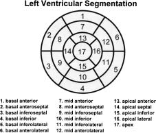
sistent, and mixed). The location of the defect must be
elements are presented in detail in Table .
described using the standardized 17-segment model
ASNC imaging guidelines for nuclear cardiology procedures
Rest Tl-201/stress Tc- 99mRest Tc-99m/stress
Viability onlyStress/rest Rb-82Stress/rest Tl-201Rest Rb-82/F-18 FDGRest/delayed restERNA in vivo labelingERNA in vitro labelingFPRNAOther
Tc-99m sestamibiTc-99m tetrofosminRb-82N-13 ammonia
Stress injection time Time of stress injection
Stress imaging time Time of stress imaging
Tc-99m sestamibiTc-99m tetrofosminRb-82N-13 ammoniaTc-99m DTPATc-99m HDP
ASNC imaging guidelines for nuclear cardiology procedures
FDG, Fluoro deoxyglucose; ERNA, equilibrium radionuclide angiocardiography; FPRNA, first-pass radionuclide angiography;SPECT, single photon emission computed tomography; DTPA, diethylene triaminepentaacetic acid; HDP, hydroxymethylenediphosphonate; PET, positron emission tomography.
(Appendix 3).The presence of apparent transient
recommended for inclusion in the report and the sug-
cavitary dilatation must be reported.
gested data elements to use when reporting quantitative
The remaining elements in Table are recom-
elements are outlined in Table . The information in
these tables may be repeated as required to describe
parameters. The use of quantitative image processing is
Basal anteroseptalBasal inferoseptalBasal inferolateralBasal anterolateralMid-anteriorMid-anteroseptalMid-inferoseptalMid-inferiorMid-inferolateral
Apical anteriorApical septalApical inferiorApical lateralApexNone
ASNC imaging guidelines for nuclear cardiology procedures
The information in this table may be repeated as required to describe multiple perfusion defects. TCD,Transient cavity dilation; TID, transient ischemic dilation; LV, left ventricular; RV, right ventricular.
SSS, Summed stress score; SRS, summed rest score; SDS, summed difference score.
ASNC imaging guidelines for nuclear cardiology procedures
Assessment of LV function and wall motion should
reported noting both their severity (i.e., hypokinesis
be performed in all patients if technically feasible with
[mild, moderate, severe], akinesis, or dyskinesis) and
stress and/or rest gated techniques. The report should
location using the standardized 17-segment model.
describe the timing of the acquisition of the image set
Optionally, LV volume data can be reported. The data
(i.e., during exercise [first pass], post-stress, or at rest),
elements specific to LV function are located in
the global LV function (i.e., normal, mild, moderate, or
Tables and . The information in these tables may
severely reduced), and the quantitative LV ejection
be repeated as required to describe multiple perfusion
fraction. Regional wall motion abnormalities should be
Mildly reducedModerately reducedSeverely reducedHyperdynamic
Mildly enlargedModerately enlargedSeverely enlarged
Mild hypokinesisModerate hypokinesisSevere hypokinesisAkinesisDyskinesis
Basal inferolateralBasal anterolateralMid-anteriorMid-anteroseptalMid-inferoseptalMid-inferiorMid-inferolateralMid-anterolateralApical anteriorApical septalApical inferiorApical lateralApexNoneDiffuse
ASNC imaging guidelines for nuclear cardiology procedures
Mildly decreased WTModerately decreased WTSeverely decreased WTHyperdynamic WT
Basal anteriorBasal anteroseptalBasal inferoseptalBasal inferolateralBasal anterolateralMid-anteriorMid-anteroseptalMid-inferoseptalMid-inferiorMid-inferolateralMid-anterolateralApical anteriorApical septalApical inferiorApical lateralApexNoneDiffuse
New functional abnormalityImprovement of function
The information in this table may be repeated as required to describe multiple perfusion defects. LV, Left ventricular; EF, ejection fraction; EDV, end-diastolic volume; ESV, end-systolic volume; WT, wall thickening.
AbnormalMildly reducedModerately reducedSeverely reduced
Mildly enlargedModerately enlargedSeverely enlarged
ASNC imaging guidelines for nuclear cardiology procedures
Mild hypokinesisModerate hypokinesisSevere hypokinesisAkinesisDyskinesis
Basal anteroseptalBasal inferoseptalBasal inferolateralBasal anterolateralMid-anteriorMid-anteroseptalMid-inferoseptalMid-inferiorMid-inferolateralMid-anterolateralApical anteriorApical septalApical inferiorApical lateralApexNoneDiffuse
Basal anteroseptalBasal inferoseptalBasal inferolateralBasal anterolateralMid-anteriorMid-anteroseptalMid-inferoseptalMid-inferior
ASNC imaging guidelines for nuclear cardiology procedures
Mid-inferolateralMid-anterolateralApical anteriorApical septalApical inferiorApical lateralApexNoneDiffuse
The information in this table may be repeated as required to describe multiple perfusion defects. LV, Left ventricular; EF, ejection fraction; FPRNA, first-pass radionuclide angiography; ERNA, equilibrium radionuclide angiocar-diography; EDV, end-diastolic volume; ESV, end-systolic volume; WT, wall thickening.
The report must also function as part of the quality
The perfusion imaging study can demonstrate
reporting mechanism for the lab. It must include a
perfusion data that allow interpretation of perfusion,
statement regarding the overall quality of the study and
size, and function of the right ventricle (RV). These may
should address the presence of potential artifacts, inci-
be optionally reported using the data elements in
dental findings, and/or extra-cardiac activity that would
Table These are usually not routinely reported
be pertinent to or limit the quality of the study. These
except in the presence of pathology or a specific indi-
cation that would warrant their inclusion in the report.
ASNC imaging guidelines for nuclear cardiology procedures
Diaphragmatic attenuationMotion artifactSubdiaphragmatic activityMisregistration artifactExtravasated doseCT for attenuation correction
Mildly reducedModerately reducedSeverely reduced
Moderate hypokinesisSevere hypokinesisAkinesisDyskinesis
RV, Right ventricular; FPRNA, first-pass radionuclide angiography; ERNA, equilibrium radionuclide angiocardiography; EF, ejectionfraction, LV, left ventricle; EDV, end-diastolic volume; ESV, end-systolic volume.
ASNC imaging guidelines for nuclear cardiology procedures
variables, however, are not covered adequately and arenot assignable to other existing tables. Table
FPRNA and ERNA utilize a number of variables
describes the variables that are specific for FPRNA and
included in other tables, such as those describing LV and
RV function at rest and with exercise. A number of
Table 14. FPRNA/ERNA (rest and exercise) specific variables
where 4 = normal,3 = mild hypokinesis,2 = moderate hypokinesis,1 = severe hypokinesis,0 = akinetic,-1 = dyskinetic
SegmentsBasal inferiorBasal anteriorBasal anteroseptalBasal inferoseptalBasal inferolateralBasal anterolateralMid-anterior
ASNC imaging guidelines for nuclear cardiology procedures
Mid-anteroseptalMid-inferoseptalMid-inferiorMid-inferolateralMid-anterolateralApical anteriorApical septalApical inferiorApical lateralApex
RA, Right atrium; LA, left atrium; LV, left ventricle; FPRNA, first-pass radionuclide angiography; ERNA, equilibrium radionuclideangiocardiography; RV, right ventricle.
metabolism mismatched defects must be described withregard to location and
Assessment of myocardial viability should include
The remaining elements in Table are recom-
visual and quantitative analysis. Metabolism defects,
mended for use in reporting myocardial viability. The
perfusion/metabolism matched defects, and perfusion/
Table 15. Viability—qualitative analysis
Basal anteriorBasal anteroseptalBasal inferoseptalBasal inferolateralBasal anterolateralMid-anteriorMid-anteroseptalMid-inferoseptal
ASNC imaging guidelines for nuclear cardiology procedures
Mid-inferolateralMid-anterolateralApical anteriorApical septalApical inferiorApical lateralApexNone
Basal anteroseptalBasal inferoseptalBasal inferolateralBasal anterolateralMid-anteriorMid-anteroseptalMid-inferoseptalMid-inferiorMid-inferolateralMid-anterolateralApical anteriorApical septalApical inferiorApical lateralApexNone
Basal anteroseptalBasal inferoseptalBasal inferolateralBasal anterolateralMid-anteriorMid-anteroseptalMid-inferoseptalMid-inferior
ASNC imaging guidelines for nuclear cardiology procedures
Mid-anterolateralApical anteriorApical septalApical inferiorApical lateralApexNone
The information in this table may be repeated as required to describe multiple metabolism defects. LV,Lleft ventricular; RV, right ventricular.
Table 16. Viability—quantitative analysis
use of quantitative image elements (i.e., number of
data elements specific to this section are outlined in
viable segments and extent of matched and mismatched
defects) is also recommended. Table outlines the
Furthermore, to ensure timely access to the data, the
quantitative data for myocardial viability.
report needs to be compliant with the standard for timely
The final component of the structured myocardial
reporting requiring completion of the interpretation
perfusion report is the most important. The Overall
within one business day and transmittal from the lab to
Impression assimilates all of the detailed findings pre-
the referring physician within two business days.
sented in the prior sections into a succinct statement
The appendices to this guideline demonstrate model
regarding whether LV perfusion and LV function are
formats for structured reporting based on the principles
normal or abnormal and describes the abnormality as
and data elements contained in this document. Appendix
ischemia or infarction. It also addresses the presence or
1 is a model format for stress myocardial perfusion
absence of significantly viable myocardium when this
imaging and Appendix 2 is a model format for phar-
macologic-based myocardial perfusion imaging. They
Additionally, the Overall Impression summarizes
are intended as examples only and ASNC fully
the stress test findings, assesses the clinical significance
acknowledges that there are many allowable structured
of the scan, and may assign vascular territories to the
formats for the reporting of nuclear myocardial perfu-
abnormalities and compare results to prior studies. The
sion images. Different structured report formats would
ASNC imaging guidelines for nuclear cardiology procedures
Left anterior descendingRight coronaryLeft circumflexLeft main
Potentially viable myocardiumNon-viable myocardium
Low riskModerate riskHigh riskUncertain risk
LV, Left ventricular; RV, right ventricular; FPRNA, first-pass radionuclide angiography; ERNA, equilibrium radionuclide angiocar-diography; MD, physician or doctor of medicine.
be required for the other indications covered in this
document (e.g., PET, exercise/rest FPRNA/ERNA, and
C. David Cooke, MSEE, receives partial royalties from the
viability imaging). Appendix 3 provides a diagram of
sale of Emory Cardiac Toolbox, and is a part-time employee of
ASNC imaging guidelines for nuclear cardiology procedures
Publication and distribution of this document are made possible
by corporate support from Astellas Pharma US, Inc; Covidien; andGE Healthcare. Corporate supporters were not involved in the
The overall quality of the study is poor/fair/good/
creation or review of information contained in this guideline.
excellent. Attenuation artifact was present/absent.
Left ventricular cavity is noted to be normal/
enlarged on the rest (and/or stress) studies. There isevidence of abnormal lung activity. Additionally, the
Stress/Rest (or Rest/Stress) Single-/Dual-
right ventricle is normal/abnormal (specify: ____).
Isotope SPECT Imaging with Exercise Stressand Gated SPECT Imaging
SPECT images demonstrate homogeneous tracer
distribution throughout the myocardium OR a small/
moderate/large perfusion abnormality of mild/mod-
(select one) Diagnosis of coronary disease
erate/severe severity is present in the ____ (location)
Evaluation of extent and severity of coronary artery
region on the stress images. The rest images reveal
____. Gated SPECT imaging reveals normal myo-
cardial thickening and wall motion. OR Gated
Risk stratification—post-myocardial infarction
SPECT imaging demonstrates hypokinesis/dyskin-
esis/akinesis of the ____ (location). The left
ventricular ejection fraction was calculated to be____% OR the left ventricular ejection fraction was
Myocardial perfusion imaging is normal/abnormal.
Previous cardiac procedures include: ____
There is a small/moderate/large area of ischemia/
infarction in the ____ location. Overall left ventric-ular systolic function was normal/abnormal with/
without regional wall motion abnormalities (as
The patient performed treadmill exercise/bicycle
noted above). Compared to the prior study from
exercise using a modified Bruce/Bruce/Naughton/
____ (date), the current study reveals ____.
____ protocol, completing ____ minutes and com-pleting an estimated workload of ____ metabolicequivalents (METS). The test was terminated due tofatigue/shortness of breath/chest pain/___. The heart
rate was ____ beats per minute at baseline and
increased to ____ beats at peak exercise, which was
____% of the maximum predicted heart rate. Therest blood pressure was ___ mm/Hg and increased/
Stress/Rest (or Rest/Stress) Single-/Dual-
decreased to ___ mm/Hg, which is a normal/
hypotensive/hypertensive response. The patient did/
did not develop any symptoms other than fatigue
during the procedure; specific symptoms include____. The resting electrocardiogram demonstrated
(select one) Diagnosis of coronary disease
____ and did/did not show ST-segment changes
Evaluation of extent and severity of coronary artery
consistent with myocardial ischemia.
Myocardial perfusion imaging was performed at rest
Risk stratification—post-MI/preoperative/general
(____ minutes following the injection of ____ mCi
of ____). At peak exercise, the patient was injected
with ____ mCi of ____ and exercise was continuedfor ____ minute(s). Gating post-stress tomographic
imaging was performed ____ minutes after stress
ASNC imaging guidelines for nuclear cardiology procedures
Previous cardiac procedures include: ____
ventricular systolic function was normal/abnormal
with/without regional wall motion abnormalities (asnoted above). Compared to the prior study from
____ (date), the current study reveals ____.
Pharmacologic stress testing was performed with
adenosine/dipyridamole/dobutamine/regadenoson ata rate of ____ for ___minutes. Additionally, low-
level exercise was performed along with the vaso-
dilator infusion (specify: ____). The heart rate was____ at baseline and rose to ____ beats per minuteduring the adenosine/dipyridamole/dobutamine/rega-denoson infusion. The rest blood pressure was ___mm/Hg and increased/decreased to ___ mm/Hg,which
response. The patient developed significant symp-toms,
electrocardiogram demonstrated ____ and did/didnot show ST-segment changes consistent with myo-cardial ischemia.
Myocardial perfusion imaging was performed at rest
(____ minutes following the injection of ____ mCiof ____). At peak pharmacologic effect, the patientwas injected with ____ mCi of ____. Gating post-stress tomographic imaging was performed ____minutes after stress (and rest).
The overall quality of the study is poor/fair/good/
excellent. Attenuation artifact was present/absent.
1. Gonzalez P, Canessa J, Massardo T. Formal aspects of the user-
Left ventricular cavity is noted to be normal/
friendly nuclear cardiology report. J Nucl Cardiol 1999;6:157.
enlarged on the rest (and/or stress) studies. There is
2. Wackers FJ. Intersocietal Commission for the Accreditation of
Nuclear Cardiology Laboratories (ICANL) position statement on
evidence of abnormal lung activity. Additionally, the
standardization and optimization of nuclear cardiology reports. J
right ventricle is normal/abnormal (specify: ____).
3. Cerqueira MD. The user-friendly nuclear cardiology report: What
SPECT images demonstrate homogeneous tracer
needs to be considered and what is included. J Nucl Cardiol
distribution throughout the myocardium OR a small/
moderate/large perfusion abnormality of mild/mod-
4. Hendel RC, Wackers FJ, Berman DS, et al. Reporting of radio-
nuclide myocardial perfusion imaging studies. J Nucl Cardiol
erate/severe severity is present in the ____ (location)
region on the stress images. The rest images reveal
5. Tilkemeier PL, Cooke CD, Ficaro EP, et al. Imaging guidelines for
____. Gated SPECT imaging reveals normal myo-
nuclear cardiology procedures: Standardized reporting of myo-
cardial thickening and wall motion. OR Gated
cardial perfusion images. J Nucl Cardiol 2009;16. doi:
SPECT imaging demonstrates hypokinesis/dyskin-
6. Digital Imaging and Communications in Medicine (DICOM).
esis/akinesis of the ____ (location). The left
Supplement 72: Echocardiography procedure reports.
ventricular ejection fraction was calculated to be
____% OR the left ventricular ejection fraction was
7. Digital Imaging and Communications in Medicine (DICOM).
Supplement 128: Cardiac stress testing structured reports.
(2009). Pub-lished 31 Oct 2008. Accessed 12 Feb 2009.
Myocardial perfusion imaging is normal/abnormal.
8. IHE. IHE technical framework volume I: Integration profiles.
There is a small/moderate/large area of ischemia/
(2009). Published 30 Aug 2007. Acessed 12 Jan 2009.
ASNC imaging guidelines for nuclear cardiology procedures
9. Douglas PS, Hendel RC, Cummings JE, et al. ACCF/ACR/AHA/
and Blood Institute and Boston University.
ASE/ASNC/HRS/NASCI/RSNA/SAIP/SCAI/SCCT/SCMR 2008
. Updated 28 Jan 2009. Accessed 19 Feb 2009.
Health policy statement on structured reporting in cardiovascular
18. Diamond GA, Hirsch M, Forrester JS, et al. Application of
imaging. J Am Coll Cardiol 2009;53:76-90.
information theory to clinical diagnostic testing. The electrocar-
10. Hendel RC, Budoff MJ, Cardella JF, et al. ACC/AHA/ACR/ASE/
diographic stress test. Circulation 1981;63:915-21.
ASNC/HRS/NASCI/RSNA/SAIP/SCAI/SCCT/SCMR/SIR
19. Mark DB, Hlatky MA, Harrel FE, et al. Exercise treadmill score
key data elements and definitions for cardiac imaging: A report of
for predicting prognosis in coronary artery disease. Ann Intern
the American College of Cardiology/American Heart Association
Task Force on Clinical Data Standards. J Am Coll Cardiol 2009;
20. Hansen CL, Goldstein RA, Akinboboye OO, et al. Imaging
guidelines for nuclear cardiology procedures: Myocardial perfu-
11. Cerqueira MD, Weissman NJ, Dilsizian V, et al. Standardized
sion and function: Single photon emission computed tomography.
myocardial segmentation and nomenclature for tomographic
imaging of the heart: A statement for healthcare professionals
21. Schoder H, Campisi R, Ohtake T, et al. Blood flow-metabolism
from the Cardiac Imaging Committee of the Council on Clinical
imaging with positron emission tomography in patients with dia-
Cardiology of the American Heart Association. J Nucl Cardiol
betes mellitus for the assessment of reversible left ventricular
contractile dysfunction. J Am Coll Cardiol 1999;33:1328-37.
12. Berman DS, Germano G. An approach to the interpretation and
22. Pagano D, Townend JN, Littler WA, et al. Coronary artery bypass
reporting of gated myocardial perfusion SPECT. In: Berman DS,
surgery as treatment for ischemic heart failure: The predictive
Germano G, editors. Clinical gated cardiac SPECT. Armonk (NY):
value of viability assessment with quantitative positron emission
tomography for symptomatic and functional outcome. J Thorac
13. Cerqueira MD. The user-friendly nuclear cardiology report: What
needs to be considered and what is included. J Nucl Cardiol
23. Gerber BL, Ordoubadi FF, Wijns W, et al. Positron emission
tomography using (18)F-fluoro-deoxyglucose and euglycaemic
14. Port SC, Berman DS, Garcia EV, et al. Imaging guidelines for
hyperinsulinaemic glucose clamp: Optimal criteria for the pre-
nuclear cardiology procedures, part 2. J Nucl Cardiol 1999;6:G47-
diction of recovery of post-ischaemic left ventricular dysfunction.
Results from the European Community Concerted Action Multi-
15. Dilsizian V, Bacharach SL, Beanlands RS, et al. Imaging guide-
center study on use of (18)F-fluoro-deoxyglucose positron
lines for nuclear cardiology procedures: PET myocardial perfusion
emission tomography for the detection of myocardial viability. Eur
and metabolism clinical imaging. J Nucl Cardiol in press.
16. Corbett JR, Akinboboye OO, Bacharach SL, et al. Imaging
24. Hendel RC, Ficaro EP, Williams KA. Timeliness of reporting
guidelines for nuclear cardiology procedures: Equilibrium radio-
results of nuclear cardiology procedures. J Nucl Cardiol
nuclide angiography. J Nucl Cardiol 2009;16. doi:
17. National Heart Lung and Blood Institute, Boston University.
Framingham heart study: A project of the National Heart, Lung
Bath Linen Bed Linen Acknowledge/Approach the Customer Build the Sale w Use open questions: who, what, when, where, w Present stock to the customer using Good, Better, Best Close the Sale w Thank you w Let me know how you get on w I Look forward to seeing you next week w Is there anything else I can help you with? Bath Linen We sell a wide assortment
THE HISTORY MAZDA MX-5 Michael R. Opalak “agile” Function: adjective Etymology: Middle French, from Latin agilis, from agere to drive, act 1 : marked by ready ability to move with quick easy grace 2 : having a quick resourceful and adaptable character 3 : quick and light in motion 4 : see Miata The Birth of a Neo-Classic • Miata introduced in fall
 ASNC imaging guidelines for nuclear cardiology procedures
Previous cardiac procedures include: ____
ventricular systolic function was normal/abnormal
with/without regional wall motion abnormalities (asnoted above). Compared to the prior study from
____ (date), the current study reveals ____.
ASNC imaging guidelines for nuclear cardiology procedures
Previous cardiac procedures include: ____
ventricular systolic function was normal/abnormal
with/without regional wall motion abnormalities (asnoted above). Compared to the prior study from
____ (date), the current study reveals ____.