RAPID COMMUNICATIONS IN MASS SPECTROMETRYRapid Commun. Mass Spectrom. 2002; 16: 2075±2082Published online in Wiley InterScience (www.interscience.wiley.com). DOI: 10.1002/rcm.828
Electrospray ionization tandem mass spectrometric study
of the aconitines in the roots of aconite
Yong Wang, Zhiqiang Liu, Fengrui Song and Shuying Liu* The New Drug Laboratory of the Changchun Institute of Applied Chemistry, Changchun 130022, P. R. China
Fragmentation pathways of aconitine-type alkaloids were investigated by electrospray ionization/ion trap multistage tandem mass spectrometry. Low-energy collision-induced dissociation ofprotonated aconitines follows a dominant first step, the elimination of the C8-substituent as aceticacid or fatty acid in MS2 spectra. Successive losses of 1±4 CH3OH molecules, 1±3 H2O, CO, benzoicacid, and CH3 or C2H5 (N-substituents) are all fragmentation pathways observed in MS3 and MS4spectra. By applying knowledge of these fragmentation pathways to the aconitines in the ethanolicextract of aconite roots, all the known aconitines were detected and also 23 unknown aconitine-typealkaloids, in which the lipo-alkaloids containing residues of 15C, 17C and 19C saturated orunsaturated fatty acids were characterized. These odd-carbon-number fatty acid substituents havenot been reported previously. Copyright # 2002 John Wiley & Sons, Ltd.
Roots of Aconite (Aconitum Carmichaeli Debx.) provide one of
teristics. Then some unknown minor lipo-alkaloids in the
the most useful herbal medicines in China. Aconitine-type
crude extract of aconite are characterized, based on these
alkaloids isolated from aconite have been shown to have
potential toxicity and wide bioactivity. Mass spectrometry(MS) methods, particularly with the development of softionization techniques, such as electrospray (ESI) and matrix-
assisted laser desorption/ionization (MALDI), have played
an important role in the characterization of isolated
Roots of aconite were purchased from a drug store.
aconitines,2±4 the quantitative analysis and the stereo-
Authentic samples of aconitine (AC), hypaconitine (HC)
chemistry of aconitines,5±7 and the direct analysis of
and mesaconitine (MA) were purchased from China Drug
aconitine mixtures. In previous work from this laboratory,
and Biological Products Inspection Institute. AC, HC and
both ESI and MALDI-MS were applied to analyze complex
MA were dissolved in chloroform to obtain a solution of
mixtures of alkaloids from aconite.8,9 However, to our
concentration of approx. 0.5 mg/mL. 8-Ole-14-benzoylmesa-
knowledge, no reports have appeared in the literature on
conine and other lipo-alkaloids were synthesized using the
the application of ESI-MS to determine the fragmentation
procedure reported by Pelletier et al.11 Aconitum alkaloids in
mechanisms of aconitines and their relationship to molecular
aconite were extracted with ethanol for 48 h at room
structure. The fragmentation pathways under low-energy
temperature, and were then analyzed by ESI-MSn.
collision-induced dissociation (CID) conditions are still notsufficiently clear to assist characterization of unknown
aconitine-type alkaloids in aconite extracts.
All experiments were performed using an LCQ ion trap
All aconitines share a common C19-norditerpenoid skele-
mass spectrometer (ThertmoFinnigan, San Jose, CA, USA)
ton. Traditionally, aconitines in crude aconite can be divided
equipped with an electrospray source and capable of
into two main types according to the substituent at position 8
analyzing ions up to m/z 2000. The spray voltage was
of the norditerpenoid skeleton: diester-diterpenoid aconi-
4.5 kV in the positive ion mode. The capillary voltage was
tines (DDAs), in which the C8 position is occupied by an
fixed at 5.0 V and capillary temperature was set 200°C.
acetyl group, and lipo-alkaloids, in which the C8 position is
Sample solutions were infused at 3 mL/min via a syringe
occupied by a fatty acid acyl group.10 The other structural
pump. Collision energies for the MSn analyses ranged from
variations lie in the substituents on the C3, C10 and N atoms
25±42% of maximum, depending on the mass of the
(Table 1). In this study we investigate the fragmentation
precursor ion; the isolation width was 2 Th.
pathways of protonated aconitines and derive the relation-ship between fragmentation behavior and structural charac-
*Correspondence to: S. Liu, The New Drug Laboratory of the
ESI-MS analysis of ethanolic extract of aconite
Changchun Institute of Applied Chemistry, Changchun 130022,
Figure 1 shows an ESI mass spectrum of the mixture of
Copyright # 2002 John Wiley & Sons, Ltd. Table 1. Structures of some known aconitum alkaloids in aconite roots
If C8 acetyl is displaced by a palmitic, linoleic or oleic acyl group, the corresponding lipo-alkaloids will be formed:
aconitines; the ions are observed in two regions, m/z 600±700
present work on fragmentations of the protonated DDAs.
and m/z 800±900. In the first region, the protonated molecules
Table 1 shows that acetyl, benzoyl, methoxy and hydroxyl
for known aconitines 1±5 were identified as m/z 616, 632, 630,
groups are possible substituents on the skeleton for all
646, and 648, respectively. In addition, a peak corresponding
DDAs, and therefore it is reasonable that neutral losses of 60,
to 10-OH-aconitine (aconifine, AF) was clearly detected at
122, 32 and 18 Da should correspond to elimination of acetic
m/z 662. In the region m/z 800±900, a set of MH ions
acid, benzoic acid, methanol and water, respectively. The
corresponding to lipo-alkaloids was observed, but their
MS2 spectra of the [M H] ions for DDAs 1±6 are
further elucidation required MSn data.
summarized in Table 2, and are discussed as follows:
(1) All fragment ions corresponding to [M H-AcOH]
provide the most abundant signals in the CID spectra,
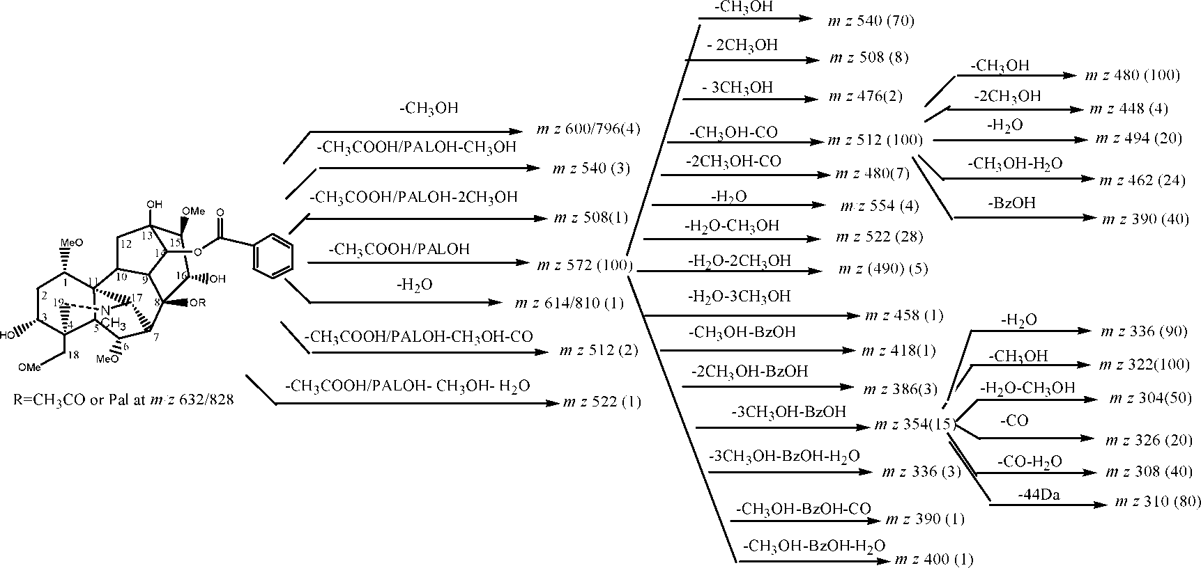
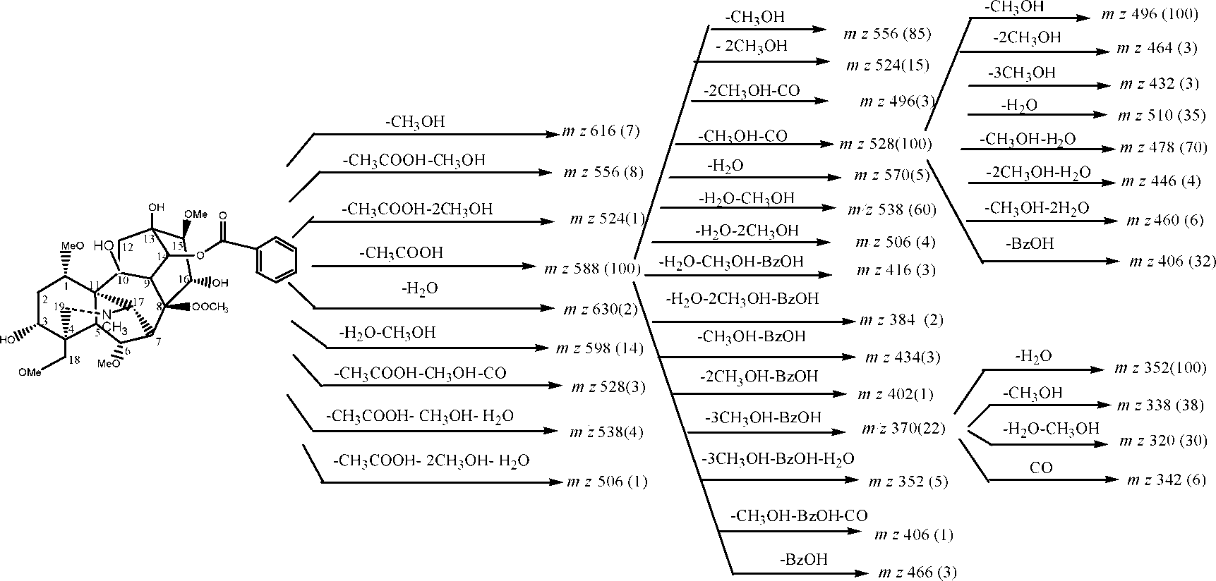
Schemes 1±3 summarize all the important findings of the
indicating that C8 is an active site.
(2) In addition to [M H-CH3OH] fragments, some very
low-abundance peaks corresponding to [M H-AcOH-CH3OH], [M H-AcOH-CH3OH-CO] and [M H-
AcOH-2CH3OH], were also detected for all DDAs.
The formation of [M H-AcOH-CH3OH-CO] sug-
gested that the C=C bond resulting from elimination ofacetic acid could be assigned to positions 8 and 15 (seeScheme 4).
(3) For DDAs 3±6, whose C3 positions are all occupied by
hydroxyl, three kinds of low-abundance fragment ions,corresponding to [M H-H2O], [M H-CH3OH-
H2O] and [M H-AcOH-CH3OH-H2O], were ob-
served; these can be viewed as characteristic fragment
Figure 1. ESI-MS of the extract from crude aconite roots.
ions for compounds with a C3 hydroxyl.
Copyright # 2002 John Wiley & Sons, Ltd.
Rapid Commun. Mass Spectrom. 2002; 16: 2075±2082
Scheme 1. Fragmentation pathways proposed according to CID spectra of HA by positive ion ESI-MS. Scheme 2. Fragmentation pathways proposed according to CID spectra of MA by positive ion ESI-MS. Scheme 3. Fragmentation pathways proposed according to CID spectra of 10-OHMA by positive ion ESI-MS.
Copyright # 2002 John Wiley & Sons, Ltd.
Rapid Commun. Mass Spectrom. 2002; 16: 2075±2082
Table 2. MS2 spectra of [M H] ions for DDAs 1–6, CID 28% Scheme 4. Proposed mechanism for successive losses of AcOH and CO from Table 3. MS3 spectra of [F1 H] = [M H À AcOH] ions, for M = DDAs 1–6; CID 27%
Copyright # 2002 John Wiley & Sons, Ltd.
Rapid Commun. Mass Spectrom. 2002; 16: 2075±2082
186] and 310 [F1 H-218-28]. It was noted that thedifferences between m/z 154, 186 and 218 are all 32 Da,corresponding to one methanol, and the difference between154 and 32 Da is 122 Da, the molecular mass of benzoic acid(BzOH). Therefore, the [F1 H] fragment at m/z 556produces successive losses of three methanol moleculesand one benzoic acid, to give a stable fragment at m/z 338. This conclusion was confirmed by the MS4 spectra of m/z 492and 460 (Fig. 2), in which ions at m/z 338 were also detected.
Structure differences were reflected in the MS3 spectra for
[M H] → [F1 H] → X. Firstly, very low-abundancefragments corresponding to loss of water were observed inthese MS3 spectra for M = HA and DA, but much moreabundant fragment ions arising from loss of water wereobserved in the MS3 spectra for M = MA, AC, BW (10-OH-MA) and AF (10-OH-AC). Secondly, [F1 H-3CH3OH-
BzOH-CO] ions were observed in these MS3 spectra forM = HA and DA, but [F1 H-3CH3OH-BzOH-H2O] were
detected instead in the MS3 spectra for M = MA, AC, BW andAF, which suggested that the C3-OH group also has a
marked effect on these MS3 spectra. In addition, reflecting
Figure 2. MS4 on (a) m/z 616[HAH] (616 >556 >492) and
the presence of the C10 hydroxyl, the peaks corresponding to
(b) m/z 616[HAH] (616 >556 >460).
simultaneous loss of methanol and water from [F1 H] forM = BW and AF were more intense than those for M = MAand AC. Finally, the abundance of [F1 H] ions alsoincreased with molecular mass, as did that of the [M H]
(4) Structural changes from N-CH3 to N-C2H5, or from H to
OH, at the C3 and C10 positions result in an increase of the
The MS4 spectra obtained for the sequence [M H] →
relative abundance of [M H]. This trend indicates that
[F1 H] →[F2 H] → X, where [F2 H] denotes
the stability of the molecular ion increased with molecu-
are summarized in Table 4. Elimination of methanol, water,
To further understand the fragmentation pathways of
benzoic acid or simultaneous losses of any one of these plus
protonated aconitines, the fragments [DDAH-AcOH],
methanol, describe all fragmentation pathways; [F2 H-
conveniently denoted [F1 H], were isolated for MS3
CH3OH] ions were observed as base peak except for
analysis (Table 3). Fragments due to loss of CO plus CH3OH
M = DA. Loss of H2O was observed in the MS4 spectra for
provide the base peak at [F1 H-CH3OH-CO] except for
M = HA and DA, suggesting that the C13 hydroxyl is
this precursor ion obtained from HA. In addition, some other
eliminated in MS4 since there is only one hydroxyl
significant fragment ions (>5%) are attributed to [F1 H-
substituent on the skeleton. In addition, presence of a
CH3OH], [F1 H-2CH3OH], [F1 H-2CH3OH-CO] and
C10-OH is reflected in the high relative abundances of
[F1 H-218]. It is somewhat difficult to explain how
[F2 H-CH3OH-H2O] in the MS4 spectra for M = BW and
[F1 H-218] ions are formed based only on these data.
AF (Table 4). Interestingly, in contrast with the above order,
Fortunately, a series of low abundance peaks (<1%) were
the fragment [DDAH-AcOH-CH3OH-CO] for DA and
also detected, which are helpful to answer this question. For
HA were detected as base peak and second strongest peak,
instance, the MS3 spectrum of the [F1 H] fragment from
respectively, suggesting that C3 being occupied by H or OH
compound HA at m/z 556 includes product ions at m/z 492
evidently influenced the stability of the [DDAH-AcOH-
[FlH-2CH3OH], 460 [FlH-3CH3OH], 478 [FlH-
CH3OH-CO-H2O], 446 [FlH-2CH3OH-CO-H2O], 432
The MS4 spectra for the sequence [M H] → [F1 H]
[FlH-3CH3OH-CO], 402 [F1 H-154], 370 [F1 H-
→ [F3 H] → X, where [F3 H] denotes the ion [M H-
Table 4. MS4 spectra for the sequence [M H]→[F1 H]→[F2 H]→X, where [F2 H] denotes M HÀAcOHÀCH3OHÀCO], for M = DDAs 1–6; CID 25%
Copyright # 2002 John Wiley & Sons, Ltd.
Rapid Commun. Mass Spectrom. 2002; 16: 2075±2082
Table 5. MS4 spectra for the sequence [M H]→[F1 H]→[F3 H]→X, where [F3 H] denotes the ion [M HÀAcOHÀ3CH3OHÀBzOH] for M = DDAs 1–6; CID 22%
AcOH-3CH3OH-BzOH], i.e. [F1 H-3CH3OH-BzOH],
behavior varies reflecting structural differences. Proposed
are listed in Table 5. Losses of CO and of CH3OH are
fragmentation pathways for [HAH], [MAH] and
common fragmentations in these MS4 experiments for all
[BWH] are shown in Schemes 2, 3 and 4, respectively.
DDAs. The odd-integer ions at m/z 295 are important becausethey indicate loss of methyl or ethyl radicals from [F3 H]
for M = HA or DA; this is followed by loss of a methanol to
Three synthesized lipo-alkaloids, including 8-pal-benzoyl-
generate a peak at m/z 263. For M = MA and AC, the
mesaconine, 8-lino-benzoylhypaconine and 8-ole-benzoyl-
distinctive reaction is loss of 44 Da, presumably CO2.
aconine, are used as examples to investigate the
The above results summarize all fragmentation pathways
fragmentation behavior of lipo-alkaloids in MS2 (Figs 3±5).
of DDAs under low-energy CID conditions. Elimination of
The fragmentation pathways of these lipo-alkaloids are the
acetic acid is the characteristic loss in MS2 for DDAs 1±6,
same as those of the corresponding acetyl compounds MA,
accompanied by losses of CH3OH and CO. Successive losses
HA and AC, except for the loss of fatty acid (256, 280, 282 Da)
of CH3OH, CO, BzOH, H2O and CH3 (N) or C2H5 (N)
rather than loss of acetic acid. It is thus understandable that
occurred in MS3 and MS4; some minor fragmentation
MS3-MS4 spectra of all fragment ions are identical with thecorresponding ions as those from the DDAs (data notshown), as summarized in Scheme 1.
ESI-MS2 for lipo-alkaloids in the extract of
The MS/MS spectra of unknown lipo-alkaloids in the aconiteextract showed interesting features compared with those ofknown lipo-alkaloids in the following two aspects:
(1) More abundant fragment ions corresponding to elimina-
tion of fatty acid were observed for the [M H] ion. Forexample, CID of m/z 866 (Fig. 1) produced two majorproduct ions at m/z 586 and 572 (Fig. 6). This is
Figure 3. MS2 spectrum on m/z 828 [8-pal-
unexpected, as we have demonstrated above that the
ion at m/z 572 should not be generated from 8-lino-benzoylaconine. The most likely simple interpretation isthat nonadecadienoic acid (MW 294 Da) was eliminated
Figure 4. MS2 spectrum on m/z 836 [8-lino- Figure 5. MS2 spectrum on m/z 868 [8-ole-
Copyright # 2002 John Wiley & Sons, Ltd.
Rapid Commun. Mass Spectrom. 2002; 16: 2075±2082
Figure 10. MS2 spectrum on m/z 826 ions of lipo-alkaloids in Figure 6. MS2 spectrum on m/z 866 ions of lipo-alkaloids in
Similarly, the MS2 spectrum of of m/z 828 (Fig. 7)
contains two major fragment ions at m/z 586 and 572,indicating losses of 242 and 256 Da, respectively, suggest-ing the presence of 8-pdc-benzoylaconine and 8-pal-
Table 6. Lipo-alkaloids from the roots of aconite characterized by Figure 7. MS2 spectrum on m/z 828 ions of lipo-alkaloids in Figure 8. MS2 spectrum on m/z 868 ions of lipo-alkaloids in Figure 9. MS2 spectrum on m/z 882 ions of lipo-alkaloids in
pdn, pdc, pme, pal, hdc, linolen, lino, ole, ndd, ndn, ecd, ecn, denote the
from the precursor ion to produce m/z 572; thus, the ion at
residues of pentadecenoic, pentadecanoic, palmitoleic, palmitic, hepta-
m/z 866 appears to correspond to two lipo-alkaloids, 8-
decanoic, linolenic, linoleic, oleic, nonadecadienoic, nonadecenoic,eicosadienoic, and eicosenoic acid, respectively
lino-benzoylaconine and 8-ndd-benzoylmesaconine.
* marks the lipo-alkaloids that have been not been reported previously in
(here ndd denotes nonadecadienoic acid).
Copyright # 2002 John Wiley & Sons, Ltd.
Rapid Commun. Mass Spectrom. 2002; 16: 2075±2082
benzoylmesaconine, respectively (here pdc and pal
denote pentadecanoic and palmitic acid, respectively). The MS2 spectrum of m/z 868 (Fig. 8) contains three major
The ESI-MSn (n = 2±4) study suggested that the neutral loss
fragment ions at m/z 588, 586 and 572, indicating losses of
of acetic acid or long-chain fatty acid is the major
280, 282 and 312 Da, respectively, suggesting the pre-
fragmentation channel in MS2 for protonated aconitines.
sence of 8-lino-10-OH-benzoylaconine, 8-ole-benzoylaco-
Further successive losses of CH3OH, H2O, CO, BzOH, and
nine and 8-arac-benzoylmesaconine, respectively (here
CH3 (N) or C2H5 (N) account for all fragmentation pathways
lino, ole and arac denote linoleic, oleic and arachidonic
in MS3 and MS4. The structural changes involving C3-OH,
C10-OH and N-CH3 or N-C2H5 can be reflected markedly in
(2) MS/MS also permits identification of minor components
ESI-MSn behavior. Based on these characteristics, ESI-MSn
of aconitine mixtures that may not be readily detected
provides a rapid and sensitive method for the direct analysis
from the ESI mass spectrum (Fig. 1) alone. For example,
of aconitine mixtures in the roots of aconite without the need
the signals at m/z 882 and 826 are very weak in Fig. 1, and
for chromatographic separation or derivatization; this
the MS2 spectra of these ions are shown in Figs 9 and 10,
advantage is most evident for differentiating the lipo-
respectively. As shown in Fig. 9, m/z 882 produced three
alkaloids with isobaric [M H] ions. Besides the pre-
main product ions at m/z 602, 586 and 572, corresponding
viously known lipo-alkaloids, a series of unknown lipo-
to losses of 280, 296 and 310 Da, respectively, suggesting
alkaloids were characterized, in which the lipo-alkaloids
that m/z 882 was composed of three different protonated
containing odd-carbon-number fatty acid residues, and
lipo-alkaloids: 8-lino-10-OH-benzoylaconine, 8-ndn-
those containing 10-OH-MA or 10-OH-AC moieties, are
benzoylaconine and 8-ecn-benzoylmesaconine (lino,
reported to be present in this plant for the first time.
ndn, ecn denote linolenic, nonadecenoic and eicosenoicacid, respectively). To our knowledge, this is the first
report of lipo-alkaloids containing 10-OH-AC and 10-
The work is supported by the National Natural Science
OH-MA in aconite. In the MS2 spectrum of m/z 826 (Fig.
10), four lipo-alkaloids, 8-pdc-benzoylaconine, 8-pme-benzoylmesaconine, 8-pal-benzoyldeoxyaconine and 8-
hdc-benzoylhypaconine, are proposed due to observa-
1. Angela A. Prog. Neurobiol. 1998; 56: 211.
tion of the fragment ions at m/z 586, 572, 570 and 556,
2. Sayed HM, Desai HK, Ross SA. J. Nat. Prod. 1992; 55: 1595.
respectively (here pdc, pme, pal, and dc denote penta-
3. Chen Y, Koelliker S, Oehme M, Katz A. J. Nat. Prod. 1999; 62:
decanoic, palmitoleic, palmitic and heptadecanoic acid,
respectively). All lipo-alkaloids observed here are ar-
4. Wang FP, Li ZB, Wang JZ. Acta Chim. Sinica (in Chinese)
ranged in Table 6 in groups that fragment by expelling a
5. Ito K, Ohyama Y, Hishinuma T. Planta Med. 1996; 62: 57.
fatty acid to yield the same product ion, i.e., the same
6. Ohta H, Seto Y, Tsunoda N. J. Chromatogr. B 1998; 714: 215.
7. Wada K, Mori T, Kawahara N. J. Mass Spectrom. 2000; 35: 435.
protonated DDA. The observation of lipo-alkaloids in
8. Sun WX, Liu SY, Liu ZQ, Song FR, Fang SP. Rapid Commun.
which C8 is occupied by a 15C, 17C or 19C saturated or
unsaturated fatty acid residue is the most significant
9. Sun WX, Song FR, Cui M, Liu SY. Planta Med. 1999; 65: 432.
finding of this study, and has not been demonstrated
10. Kitagawa I, Yoshikawa M, Chen ZL, Kobayashi K. Chem.
11. Bai Yl, Desai HK, Pelletier SW. J. Nat. Prod. 1994; 57: 963.
Copyright # 2002 John Wiley & Sons, Ltd.
Rapid Commun. Mass Spectrom. 2002; 16: 2075±2082
Institut de Formation en Soins Infirmiers 3ème année Promotion : 2009-2012 EVALUATION 4.7 S 5 SOINS PALLIATIFS ET DE FIN DE VIE Le 04/11/2011 REMARQUE : des informations complémentaires sont apportées pour certaines thérapeutiques et la pathologie (identifiées par *) et figurent après la situation. Mr Z., 58 ans est hospitalisé en juin 2009 ; il est alors po
How do I know whether I am at risk of having thyroid The thyroid is a butterfly-shaped gland, about the hormonal imbalance and how can it be diagnosed?size of two thumbs, located in front of the High or low thyroid hormone levels can be easily diagnosed through a blood test. windpipe. Its main function is to produce, store
 RAPID COMMUNICATIONS IN MASS SPECTROMETRYRapid Commun. Mass Spectrom. 2002; 16: 2075±2082Published online in Wiley InterScience (www.interscience.wiley.com). DOI: 10.1002/rcm.828
Electrospray ionization tandem mass spectrometric study
of the aconitines in the roots of aconite
Yong Wang, Zhiqiang Liu, Fengrui Song and Shuying Liu*
RAPID COMMUNICATIONS IN MASS SPECTROMETRYRapid Commun. Mass Spectrom. 2002; 16: 2075±2082Published online in Wiley InterScience (www.interscience.wiley.com). DOI: 10.1002/rcm.828
Electrospray ionization tandem mass spectrometric study
of the aconitines in the roots of aconite
Yong Wang, Zhiqiang Liu, Fengrui Song and Shuying Liu*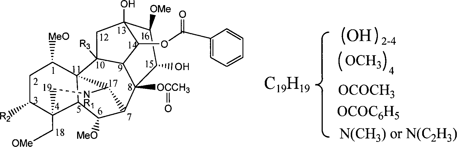

 Table 1. Structures of some known aconitum alkaloids in aconite roots
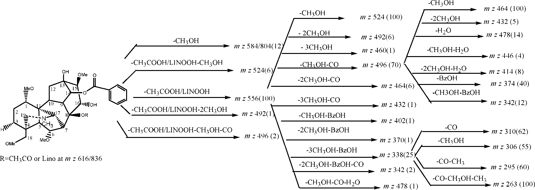
 Scheme 1. Fragmentation pathways proposed according to CID spectra of HA by positive ion ESI-MS.
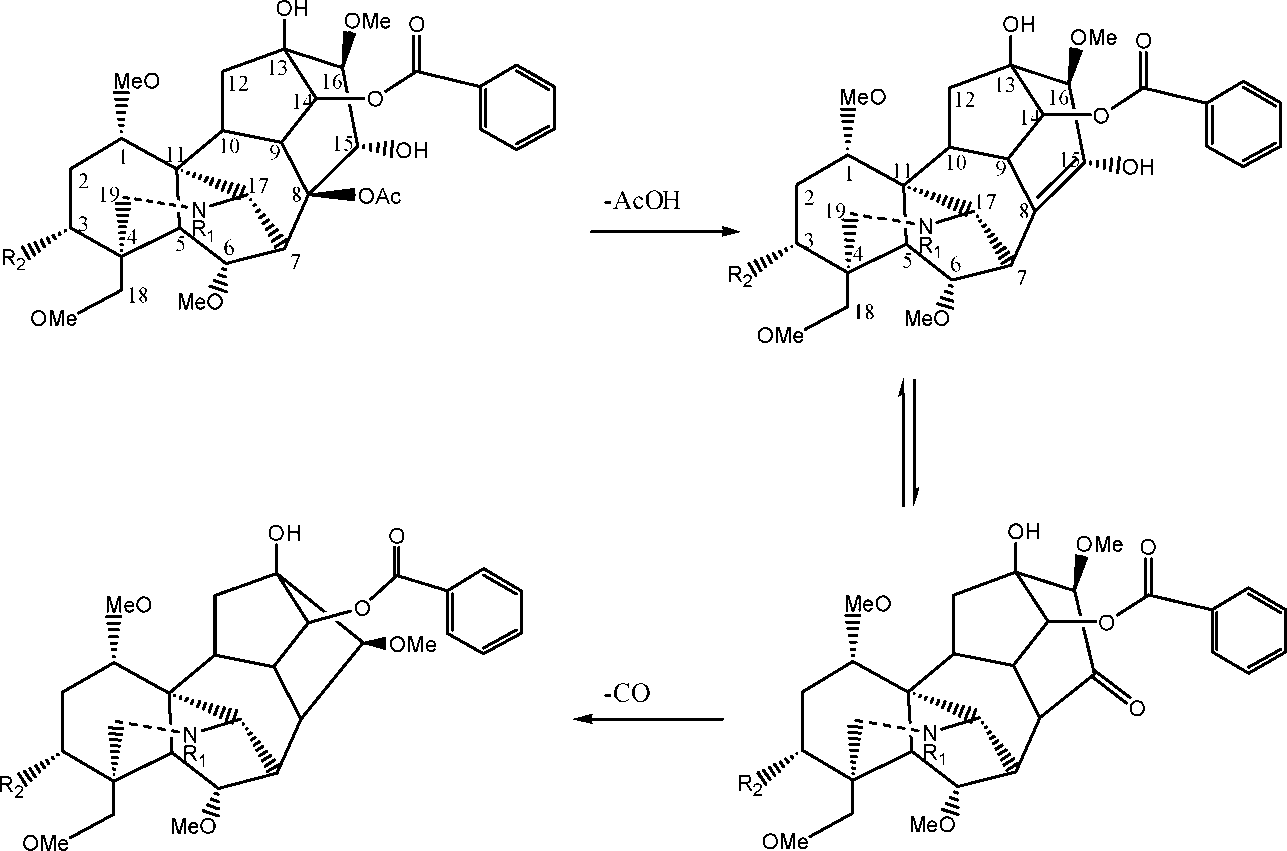
 Table 2. MS2 spectra of [M H] ions for DDAs 1–6, CID 28%
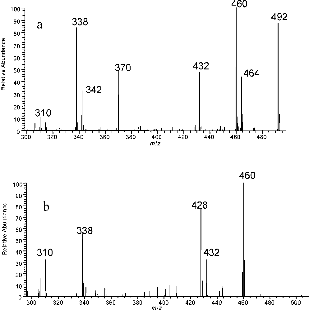
 186] and 310 [F1 H-218-28]. It was noted that thedifferences between m/z 154, 186 and 218 are all 32 Da,corresponding to one methanol, and the difference between154 and 32 Da is 122 Da, the molecular mass of benzoic acid(BzOH). Therefore, the [F1 H] fragment at m/z 556produces successive losses of three methanol moleculesand one benzoic acid, to give a stable fragment at m/z 338.
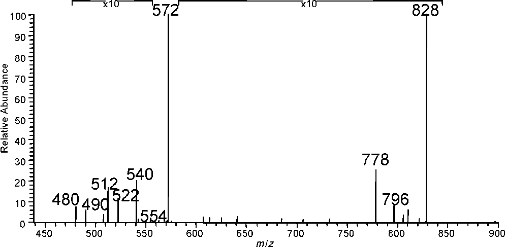
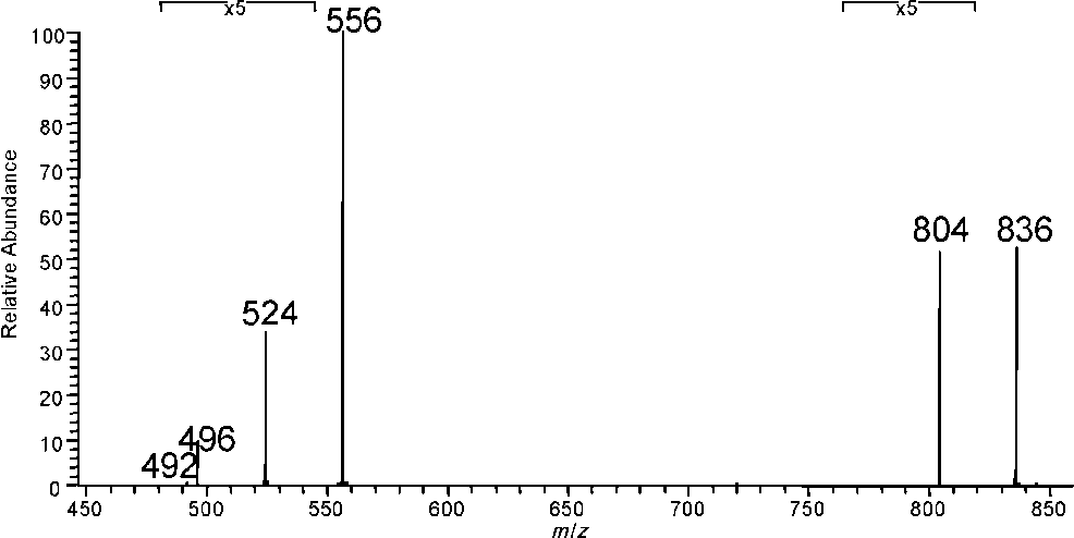
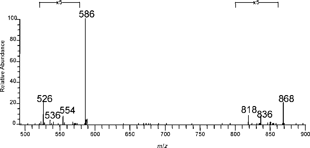
 Table 5. MS4 spectra for the sequence [M H]→[F1 H]→[F3 H]→X, where [F3 H] denotes the ion
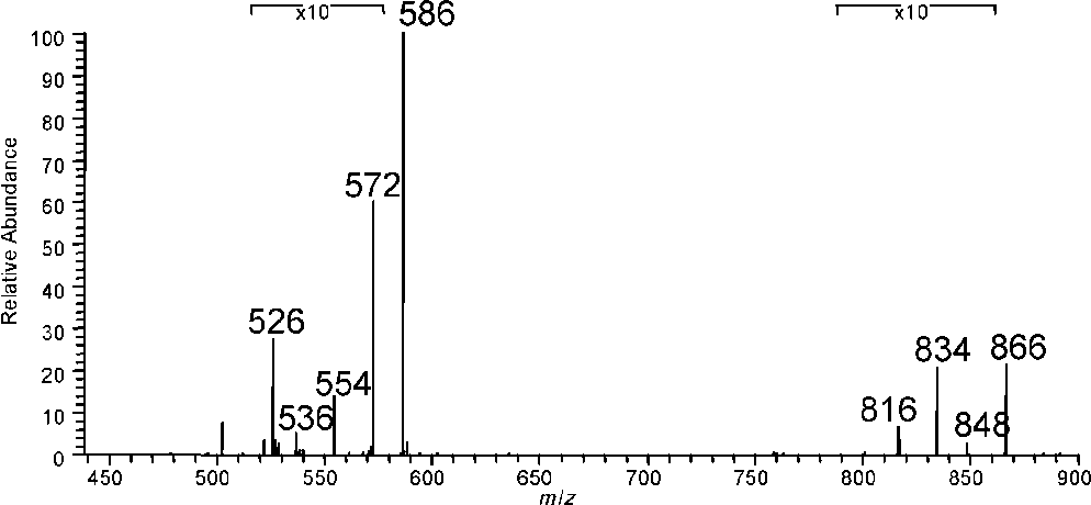
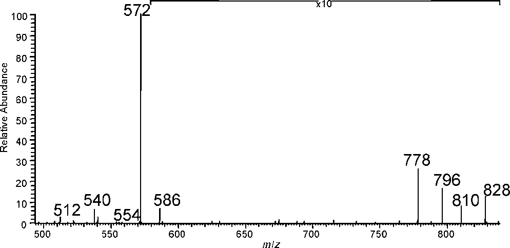
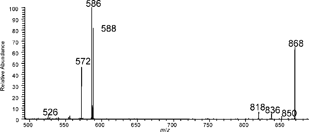
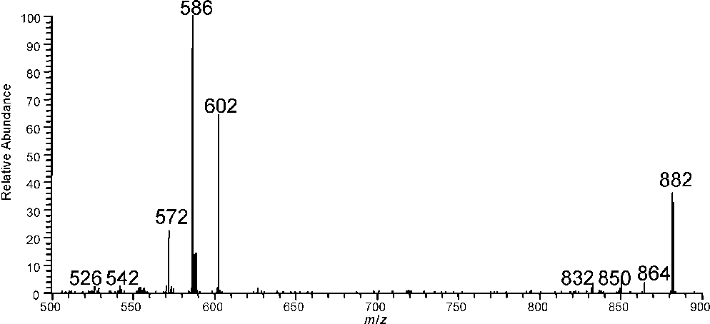
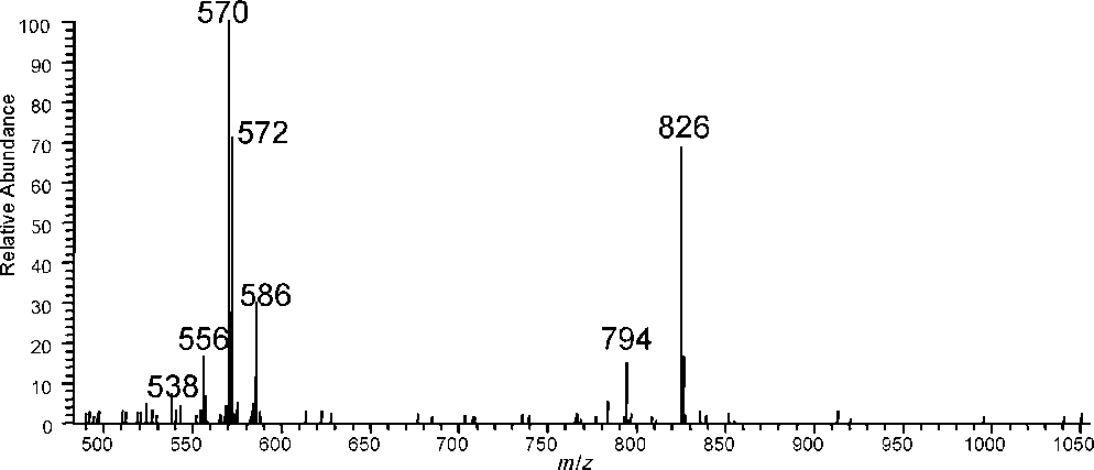
 Figure 10. MS2 spectrum on m/z 826 ions of lipo-alkaloids in
 benzoylmesaconine, respectively (here pdc and pal
denote pentadecanoic and palmitic acid, respectively).
benzoylmesaconine, respectively (here pdc and pal
denote pentadecanoic and palmitic acid, respectively).